
Immortalized Rat Hepatic Stellate Cells (CFSC-8B)
Cat.No.: CSC-I2672Z
Culture Properties: Adherent
- Specification
- Background
- Scientific Data
- Q & A
- Customer Review
The CFSC-8B cell line is an immortalized rat hepatic stellate cell (HSC) line derived from the liver of a rat with carbon tetrachloride (CCl₄)-induced cirrhosis, first established in 1991. Like other HSCs, these perisinusoidal cells reside in the space of Disse and play a central role in maintaining the hepatic microenvironment, primarily storing vitamin A in their lipid droplets during quiescence. In response to chronic liver injury (or standard culture on plastic), HSCs activate and transdifferentiate into a myofibroblast-like state capable of producing and secreting large quantities of extracellular matrix (ECM) compounds, notably type I collagen, which are the hallmark of hepatic fibrosis.
The CFSC-8B cell line exhibits a proliferating myofibroblast-like phenotype expressing key markers and cytokines associated with fibrogenesis. The cells express α-smooth muscle actin (α-SMA), desmin, glial fibrillary acidic protein (GFAP), collagens (types I and III), and soluble mediators including transforming growth factor-beta (TGF-β), platelet-derived growth factor (PDGF), and hepatocyte growth factor (HGF). They also contain abundant lipid droplets and Golgi apparatus, functional scavenger receptors (e.g., Stabilin‑1, LRP‑1), and respond to exogenous TGF‑β1 or acetaldehyde activation by further upregulating ECM production and proliferation.
As with other CFSC derivatives, the CFSC-8B genome exhibits extensive chromosome rearrangements and copy‑number variations (pseudotriploid karyotype), which should be considered when interpreting results concerning genetic stability. However, the major advantage of this line lies in its homogeneous, reproducible, and unlimited supply, overcoming the substantial hurdles of primary HSC isolation, which requires specialized equipment, trained personnel, and regulatory approval. Consequently, CFSC‑8B is an essential tool for high‑throughput antifibrotic drug screening, mechanistic studies of the TGF‑β1/Smad signaling axis in fibrosis, and investigations of liver regeneration and toxicology.
Insulin-like Growth Factor-I Reduces Collagen Production by Hepatic Stellate Cells
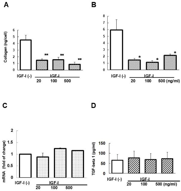
The liver is the major source of circulating insulin-like growth factor (IGF)-I. Serum IGF-I levels are decreased in cirrhotic patients depending on severity. IGF-I administration was shown to improve liver function in patients and animal models of liver cirrhosis. The effects of IGF-I on collagen accumulation by hepatic stellate cells (HSCs) and its mechanisms were studied.
A moderately activated HSC clone (CFSC-8B) was used to determine the effect of IGF-I administration on the collagen production system, including its degradation. The intracellular signaling system was also studied in the cells stimulated by IGF-I.
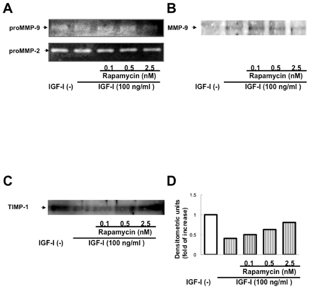
IGF-I treatment reduced total amounts of collagen deposition in a dose-related manner, while DNA synthesis was stimulated by IGF-I. IGF-I treatment did not affect transforming growth factor-beta levels and type I procollagen mRNA expression. Expression of matrix metalloproteinase (MMP)-2 and -9 was upregulated, and tissue inhibitor of metalloproteinase (TIMP)-1 expression was downregulated by IGF-I treatment. Rapamycin, an inhibitor of mammalian target of rapamycin (mTOR), suppressed phosphorylation of 70 kDa ribosomal protein S6 kinase and eukaryotic initiation factor 4E-binding protein 1, and abrogated IGF-I-induced increase in MMP-2 and -9 expression and decrease in TIMP-1 expression.
Ask a Question
Write your own review
Description: The Immortalized Mouse Prostate Epithelial Cells (iMPEC, clone7) is derived by co-expression of adenovirus E1A and dominant-negative p53 (p53DD). In addition to its non-tumorigenicity, this cell line ...

Description: Immortalized Mouse Primary Artery Fibroblasts-GFP provided by Creative Bioarray have been developed by immortalizing mouse artery fibroblasts with SV40 Large T antigen and transfecting with tGFP. The ...
Description: Immortalized MHC II -/- Mouse Macrophage Cells (C2D) is derived from the knockout mice negative for MHCII molecule. They are stable in culture and survived through crisis in which they spontaneously ...

Description: The bone morphogenetic proteins (BMPs) produced by calvarial cells are crucial in osteoblastic differentiation and bone regeneration, however the limited lifespan of primary cells diminishes their ...
Description: The Immortalized Mouse Atrioventricular Cushion Mesenchymal Cells (tsA58-AVM) were derived from atrioventricular (AV) cushions of H-2Kb -tsA58 embryos at E9.5 and were conditionally immortalized in ...
Description: IDG-SW3 represent a non-homogenous population progressing from early osteoblasts to late osteocytic. These cells express functional SV40 large T antigen that is induced in the presence of IFN γ under ...
- Adipose Tissue-Derived Stem Cells
- Human Neurons
- Mouse Probe
- Whole Chromosome Painting Probes
- Hepatic Cells
- Renal Cells
- In Vitro ADME Kits
- Tissue Microarray
- Tissue Blocks
- Tissue Sections
- FFPE Cell Pellet
- Probe
- Centromere Probes
- Telomere Probes
- Satellite Enumeration Probes
- Subtelomere Specific Probes
- Bacterial Probes
- ISH/FISH Probes
- Exosome Isolation Kit
- Human Adult Stem Cells
- Mouse Stem Cells
- iPSCs
- Mouse Embryonic Stem Cells
- iPSC Differentiation Kits
- Mesenchymal Stem Cells
- Immortalized Human Cells
- Immortalized Murine Cells
- Cell Immortalization Kit
- Adipose Cells
- Cardiac Cells
- Dermal Cells
- Epidermal Cells
- Peripheral Blood Mononuclear Cells
- Umbilical Cord Cells
- Monkey Primary Cells
- Mouse Primary Cells
- Breast Tumor Cells
- Colorectal Tumor Cells
- Esophageal Tumor Cells
- Lung Tumor Cells
- Leukemia/Lymphoma/Myeloma Cells
- Ovarian Tumor Cells
- Pancreatic Tumor Cells
- Mouse Tumor Cells