
Immortalized Human Umbilical Vein Endothelial Cells (SV40)-GFP
Cat.No.: CSC-I2045Z
Species: homo sapiens
Morphology: Polygonal
Culture Properties: Adherent
- Specification
- Background
- Scientific Data
- Q & A
- Customer Review
- Documents
free from contaminations (bacteria incl. mycoplasma, fungi, HIV, HAV, HBV, HCV, Parvo-B19) and cross-contaminations
Note: Never can cells be kept at -20°C.
Immortalized human umbilical vein endothelial cells (SV40)-GFP (HUVEC/SV40-GFP) are generated by introducing the SV40 large T antigen into primary HUVECs, followed by stable expression of green fluorescent protein (GFP). The SV40 T antigen confers indefinite proliferative capacity, while GFP provides a constitutive, non-invasive fluorescent label. Together, these features yield a scalable, trackable, and reproducible endothelial model.
The cells typically retain core endothelial markers, including CD31/PECAM-1, VE-cadherin, VEGFR2, CD34, von Willebrand factor, and eNOS. Functionally, they respond to VEGF and inflammatory cytokines and support proliferation, migration, tube formation, acetylated LDL uptake, barrier assays, leukocyte adhesion, and nitric oxide signaling. GFP expression enables real-time visualization, fluorescence-activated cell sorting, accurate quantification, and live-cell tracking in co-culture and 3D systems.
Compared with primary HUVECs, HUVEC/SV40-GFP overcome limited lifespan and donor variability, providing continuous supply, high batch consistency, and cost-effective expansion for high-throughput screening, siRNA/CRISPR perturbation, and long-term studies. They are widely used in angiogenesis, vascular permeability, inflammation, atherosclerosis, thrombosis, and endothelial dysfunction research, as well as drug discovery, target validation, toxicity testing, and host–pathogen studies. GFP labeling further facilitates co-culture with tumor cells, immune cells, or fibroblasts and supports imaging-based assays.
Uratylated Albumin Activates Endothelial Cells to Induce Monocyte Adhesion and The Release of Pro-Inflammatory Cytokines
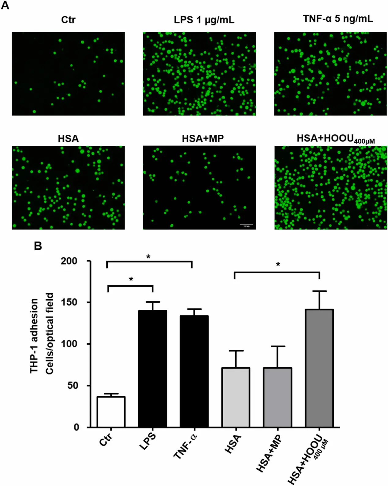
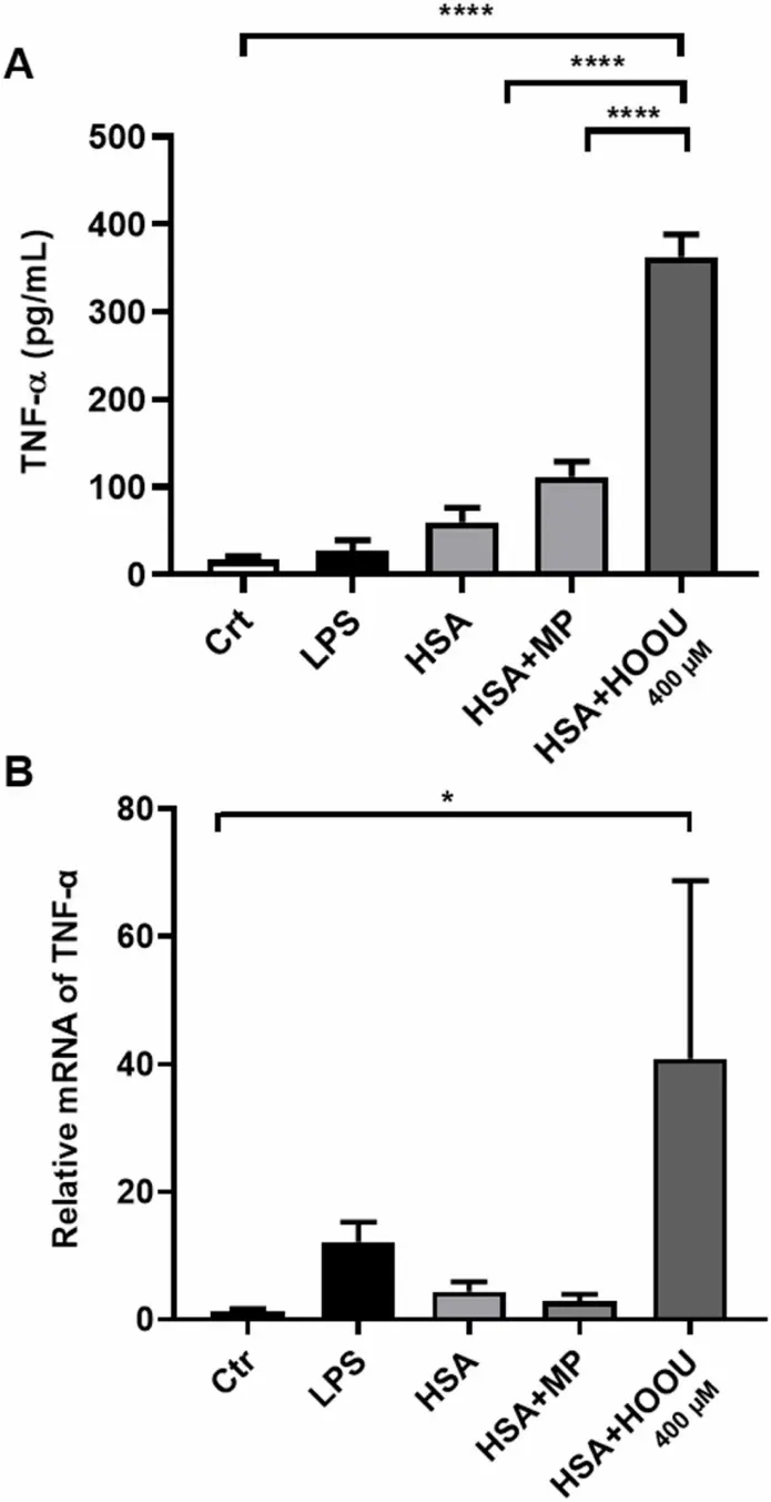
Previous studies have demonstrated a link between uric acid (UA) oxidation products, particularly urate hydroperoxide (HOOU), and albumin modifications through a process termed uratylation. Therefore, the aim of this study was to investigate the structural and functional consequences of uratylation on albumin and its effects on endothelial cells. Uratylation promotes a decrease in enthalpy change of denaturation of albumin and binding of Anilino-Naphthalene-Sulfonic acid (ANS), suggesting slight changes in protein structure. Uratylated albumin reduced human umbilical vein endothelial cell (HUVEC) migration, increased intercellular adhesion molecule-1 (ICAM-1) expression and, consequently, monocyte adhesion (THP-1), indicating endothelial activation. Furthermore, the presence of uratylated albumin stimulated the release of tumor necrosis factor-α (TNF-α). These findings provide novel insights into the mechanistic pathways connecting uric acid oxidation, post-translational modification on albumin and inflammation, highlighting the potential role of uratylated albumin in endothelial dysfunction and atherogenesis.
Ask a Question
Write your own review
 Immortalized Human Umbilical Vein Endothelial Cells (SV40)-GFP
Description: Immortalized Human Gastric Epithelial Cells-SV40 have been obtained immortalizing Human Gastric Epithelial Cells with Lenti-SV40 Lentivirus. Immortalized cells were controlled passaging side by side ...
Description: Immortalized Human Kidney Podocytes-SV40 have been obtained immortalizing Human Kidney Podocytes with Lenti-SV40 Lentivirus. Immortalized cells were controlled passaging side by side with the primary ...
Description: Nasal epithelial cells form the outermost protective layer against environmental factors. They clean, humidify, and warm inhaled air. They produces mucus, which binds particles that are subsequently ...
Description: Immortalized Human Splenic Endothelial Cells-SV40 have been obtained immortalizing Human Splenic Endothelial Cells with SV40LT expressing lentiviral particles. Immortalized cells were controlled ...
Description: Immortalized Human Corneal Epithelial Cells-SV40 have been obtained immortalizing Human Corneal Epithelial Cells with Lenti-SV40 Lentivirus. Immortalized cells were controlled passaging side by side ...
- Adipose Tissue-Derived Stem Cells
- Human Neurons
- Mouse Probe
- Whole Chromosome Painting Probes
- Hepatic Cells
- Renal Cells
- In Vitro ADME Kits
- Tissue Microarray
- Tissue Blocks
- Tissue Sections
- FFPE Cell Pellet
- Probe
- Centromere Probes
- Telomere Probes
- Satellite Enumeration Probes
- Subtelomere Specific Probes
- Bacterial Probes
- ISH/FISH Probes
- Exosome Isolation Kit
- Human Adult Stem Cells
- Mouse Stem Cells
- iPSCs
- Mouse Embryonic Stem Cells
- iPSC Differentiation Kits
- Mesenchymal Stem Cells
- Immortalized Human Cells
- Immortalized Murine Cells
- Cell Immortalization Kit
- Adipose Cells
- Cardiac Cells
- Dermal Cells
- Epidermal Cells
- Peripheral Blood Mononuclear Cells
- Umbilical Cord Cells
- Monkey Primary Cells
- Mouse Primary Cells
- Breast Tumor Cells
- Colorectal Tumor Cells
- Esophageal Tumor Cells
- Lung Tumor Cells
- Leukemia/Lymphoma/Myeloma Cells
- Ovarian Tumor Cells
- Pancreatic Tumor Cells
- Mouse Tumor Cells