
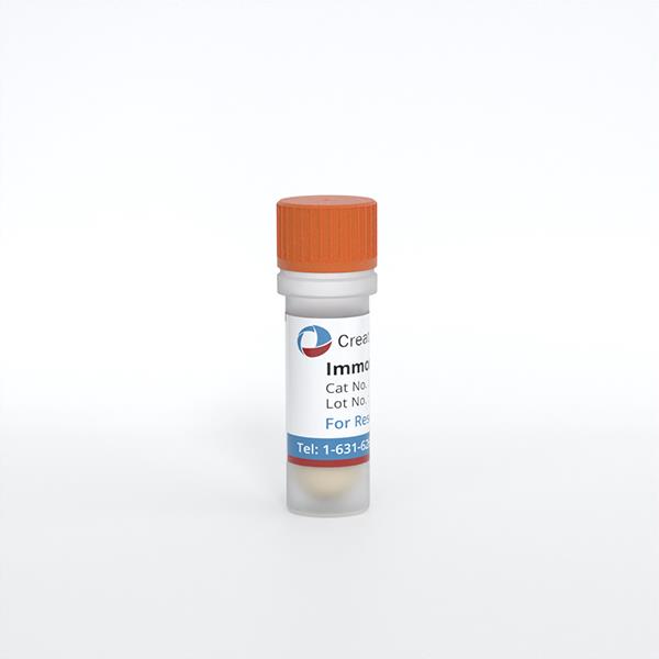
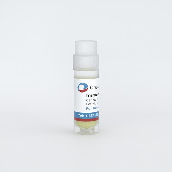
Immortalized Human Small Airway Epithelial Cells-SV40
Cat.No.: CSC-I9068L
Species: Homo sapiens
Source: Lung
Morphology: Polygonal
Culture Properties: Adherent
- Specification
- Background
- Scientific Data
- Q & A
- Customer Review
Note: Never can cells be kept at -20 °C.
CIK-HT003 HT® Lenti-SV40T Immortalization Kit
Immortalized human small airway epithelial cells-SV40 (HSAEC/SV40) are generated by introducing the SV40 large T antigen into primary human small airway epithelial cells. This confers indefinite proliferative capacity while preserving core epithelial features, including pan-cytokeratin, E-cadherin, ZO-1, and occludin. Depending on the line, they may retain club cell-associated markers such as SCGB1A1/CC10 and mucus-related proteins. Functionally, they form barrier-competent monolayers and respond to inflammatory and microbial stimuli via Toll-like receptors, secreting cytokines and chemokines such as IL-6, IL-8, and TNF-α.
Compared with primary HSAECs, HSAEC/SV40 offer a continuous, scalable, and cost-effective source with high batch-to-batch consistency and reduced donor variability. This enables reproducible high-throughput screening, long-term culture, and gene perturbation using siRNA or CRISPR. They are widely used to model small airway epithelial injury and inflammation in COPD, asthma, cystic fibrosis, and respiratory viral infections, including RSV, influenza, and SARS-CoV-2. They also support inhalation toxicology, drug transport, and biocompatibility testing.
PIWIL4 Regulates Gene Expression and piRNA Levels in RSV-Infected Airway Epithelial Cells
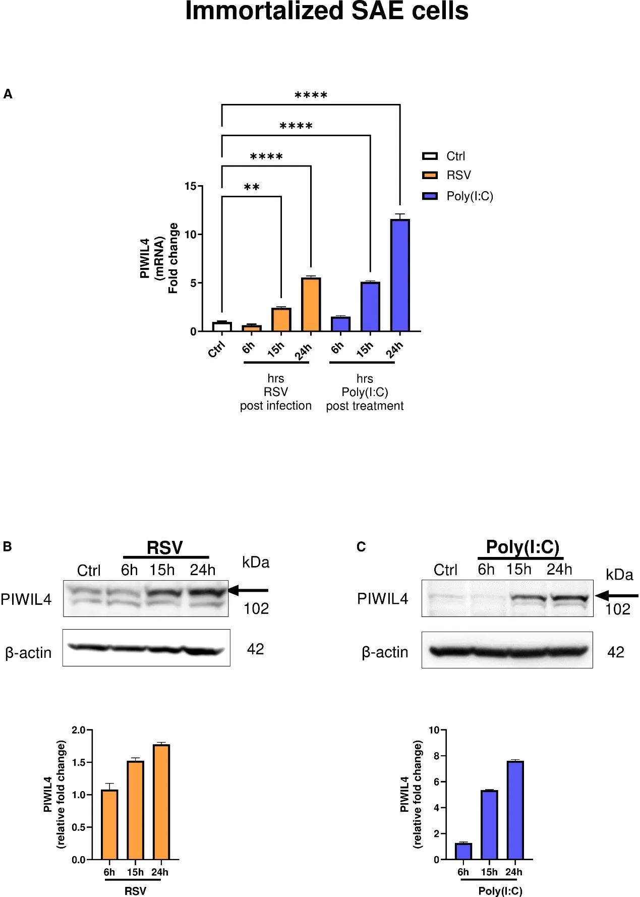
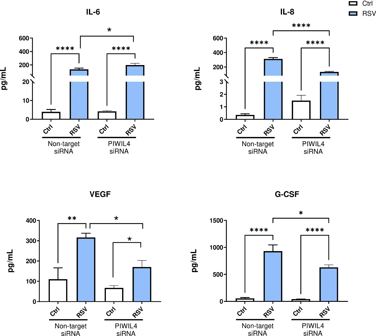
Respiratory syncytial virus (RSV) causes acute lower respiratory tract infection in young children, the elderly, and immunocompromised hosts, and no RSV vaccine or therapy exists for infants, beyond prophylaxis. PIWI-interacting RNAs (piRNAs), small non-coding RNAs acting through P element-induced wimpy testis (Piwi) proteins, remain largely unexplored. This study characterized PIWIL4 expressions and function during RSV infection or poly(I:C) stimulation, a proxy of viral infection. PIWIL4 was expressed in primary and immortalized small airway epithelial cells following RSV infection. While PIWIL4 knockdown did not affect RSV replication, it decreased cytokine and growth factor secretion, altered gene and piRNA expression, and modulated pathways involved in cytokine production, metabolism, and airway remodeling. Limited overlap between piRNA targets and transcriptomic changes suggests PIWIL4 regulates airway epithelial cell responses partly independently of piRNAs. This study positions PIWIL4 as a key regulator of airway epithelial cell responses to viral infections.
Ask a Question
Write your own review
Description: Immortalized Human Gastric Epithelial Cells-SV40 have been obtained immortalizing Human Gastric Epithelial Cells with Lenti-SV40 Lentivirus. Immortalized cells were controlled passaging side by side ...
Description: Immortalized Human Kidney Podocytes-SV40 have been obtained immortalizing Human Kidney Podocytes with Lenti-SV40 Lentivirus. Immortalized cells were controlled passaging side by side with the primary ...
Description: Nasal epithelial cells form the outermost protective layer against environmental factors. They clean, humidify, and warm inhaled air. They produces mucus, which binds particles that are subsequently ...
Description: Immortalized Human Splenic Endothelial Cells-SV40 have been obtained immortalizing Human Splenic Endothelial Cells with SV40LT expressing lentiviral particles. Immortalized cells were controlled ...
Description: Immortalized Human Corneal Epithelial Cells-SV40 have been obtained immortalizing Human Corneal Epithelial Cells with Lenti-SV40 Lentivirus. Immortalized cells were controlled passaging side by side ...
- Adipose Tissue-Derived Stem Cells
- Human Neurons
- Mouse Probe
- Whole Chromosome Painting Probes
- Hepatic Cells
- Renal Cells
- In Vitro ADME Kits
- Tissue Microarray
- Tissue Blocks
- Tissue Sections
- FFPE Cell Pellet
- Probe
- Centromere Probes
- Telomere Probes
- Satellite Enumeration Probes
- Subtelomere Specific Probes
- Bacterial Probes
- ISH/FISH Probes
- Exosome Isolation Kit
- Human Adult Stem Cells
- Mouse Stem Cells
- iPSCs
- Mouse Embryonic Stem Cells
- iPSC Differentiation Kits
- Mesenchymal Stem Cells
- Immortalized Human Cells
- Immortalized Murine Cells
- Cell Immortalization Kit
- Adipose Cells
- Cardiac Cells
- Dermal Cells
- Epidermal Cells
- Peripheral Blood Mononuclear Cells
- Umbilical Cord Cells
- Monkey Primary Cells
- Mouse Primary Cells
- Breast Tumor Cells
- Colorectal Tumor Cells
- Esophageal Tumor Cells
- Lung Tumor Cells
- Leukemia/Lymphoma/Myeloma Cells
- Ovarian Tumor Cells
- Pancreatic Tumor Cells
- Mouse Tumor Cells