
Immortalized Human Retinal Microvascular Endothelial Cells-SV40T
Cat.No.: CSC-I2309Z
Species: homo sapiens
Morphology: Polygonal
Culture Properties: Adherent
- Specification
- Background
- Scientific Data
- Q & A
- Customer Review
free from contaminations (bacteria incl. mycoplasma, fungi, HIV, HAV, HBV, HCV, Parvo-B19) and cross-contaminations
Immortalized human retinal microvascular endothelial cells-SV40T (HRMEC/SV40T) are generated by introducing the SV40 large T antigen into primary human retinal microvascular endothelial cells. This confers indefinite proliferative capacity while preserving core endothelial features, including CD31/PECAM-1, VE-cadherin, VEGFR2, and CD34, as well as blood-retinal barrier (BRB)-associated junctional proteins such as claudin-5, occludin, and ZO-1. Functionally, they respond to VEGF and inflammatory stimuli and support proliferation, migration, tube formation, and barrier assays.
Compared with primary HRMECs, HRMEC/SV40T offer a continuous, scalable, and cost-effective source with high batch-to-batch consistency and reduced donor variability. This enables reproducible high-throughput screening, gene perturbation (siRNA/CRISPR), and long-term studies. They are widely used to model retinal angiogenesis and BRB dysfunction in diabetic retinopathy, retinopathy of prematurity, retinal vein occlusion, and macular edema, and to evaluate anti-VEGF agents, corticosteroids, and metabolic or inflammatory modulators.
Limitations: SV40 large T antigen inactivates p53 and Rb and may alter stress/apoptosis responses, barrier tightness, and karyotype. Results should therefore be validated against low-passage primary HRMECs when physiological fidelity is critical. Overall, HRMEC/SV40T provide a practical, human-relevant, and reproducible platform for retinal vascular biology and translational drug discovery.
Involvement of the miR-128-3p in High Glucose-Induced Inflammatory Injury in Retinal Endothelial Cells
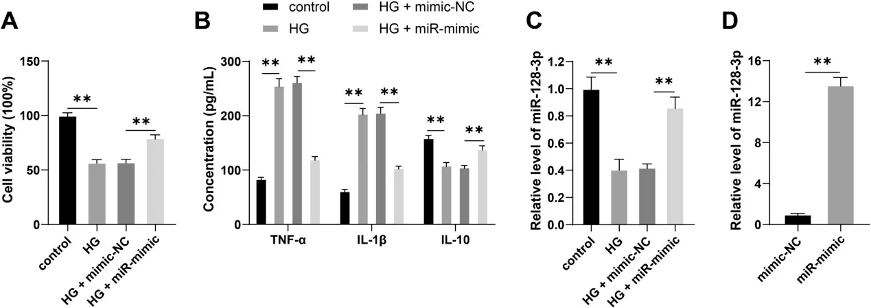
This study explores the regulatory mechanism of the miR-128-3p in diabetic retinopathy (DR)-associated inflammatory injury. A cellular model of DR was established by inducing immortalized human retinal endothelial cells (IM-HRECs) with high-glucose (HG). Cell viability was evaluated by CCK-8 assay, and the levels of TNF-α, IL-1β, and IL-10 were measured by ELISA. RT-qPCR was performed to determine miR-128-3p expression, and miR-128-3p mimics were transfected into cells to verify its regulatory role in DR-associated inflammatory injury. The results revealed that HG treatment significantly reduced both IM-HREC viability and IL-10 levels, increased the levels of TNF-α and IL-1β, and downregulated the expression of miR-128-3p. Overexpression of miR-128-3p reduced inflammation in IM-HRECs induced by HG.
Ask a Question
Write your own review

Description: Immortalized Human Gastric Epithelial Cells-SV40 have been obtained immortalizing Human Gastric Epithelial Cells with Lenti-SV40 Lentivirus. Immortalized cells were controlled passaging side by side ...
Description: Immortalized Human Kidney Podocytes-SV40 have been obtained immortalizing Human Kidney Podocytes with Lenti-SV40 Lentivirus. Immortalized cells were controlled passaging side by side with the primary ...
Description: Nasal epithelial cells form the outermost protective layer against environmental factors. They clean, humidify, and warm inhaled air. They produces mucus, which binds particles that are subsequently ...
Description: Immortalized Human Splenic Endothelial Cells-SV40 have been obtained immortalizing Human Splenic Endothelial Cells with SV40LT expressing lentiviral particles. Immortalized cells were controlled ...
Description: Immortalized Human Corneal Epithelial Cells-SV40 have been obtained immortalizing Human Corneal Epithelial Cells with Lenti-SV40 Lentivirus. Immortalized cells were controlled passaging side by side ...
- Adipose Tissue-Derived Stem Cells
- Human Neurons
- Mouse Probe
- Whole Chromosome Painting Probes
- Hepatic Cells
- Renal Cells
- In Vitro ADME Kits
- Tissue Microarray
- Tissue Blocks
- Tissue Sections
- FFPE Cell Pellet
- Probe
- Centromere Probes
- Telomere Probes
- Satellite Enumeration Probes
- Subtelomere Specific Probes
- Bacterial Probes
- ISH/FISH Probes
- Exosome Isolation Kit
- Human Adult Stem Cells
- Mouse Stem Cells
- iPSCs
- Mouse Embryonic Stem Cells
- iPSC Differentiation Kits
- Mesenchymal Stem Cells
- Immortalized Human Cells
- Immortalized Murine Cells
- Cell Immortalization Kit
- Adipose Cells
- Cardiac Cells
- Dermal Cells
- Epidermal Cells
- Peripheral Blood Mononuclear Cells
- Umbilical Cord Cells
- Monkey Primary Cells
- Mouse Primary Cells
- Breast Tumor Cells
- Colorectal Tumor Cells
- Esophageal Tumor Cells
- Lung Tumor Cells
- Leukemia/Lymphoma/Myeloma Cells
- Ovarian Tumor Cells
- Pancreatic Tumor Cells
- Mouse Tumor Cells