

Immortalized Human Pancreatic Duct Epithelial Cells-HPV16 E6/E7
Cat.No.: CSC-I9013L
Species: Homo sapiens
Source: Pancreatic Duct
Morphology: Polygonal
Culture Properties: Adherent
- Specification
- Background
- Scientific Data
- Q & A
- Customer Review
Note: Never can cells be kept at -20 °C.
Immortalized human pancreatic duct epithelial cells generated via HPV16 E6/E7 gene transduction, including the well-characterized HPDE6-E6E7 and HPDE-4/E6E7 lines, are powerful in vitro models for studying pancreatic duct biology and carcinogenesis. These cells were established using retroviral transduction of the HPV16 E6 and E7 genes into primary human pancreatic duct epithelial cells derived from normal or benign adult pancreas.
The key advantage of these cells is their near-normal genotype and phenotype, despite the targeted disruption of the p53 and pRB pathways by E6 and E7, respectively. Specifically, they retain normal genotypes for critical oncogenes and tumor suppressors, including Ki-ras, c-myc, and p16INK4A. They express key ductal epithelial markers such as carbonic anhydrase II, MUC-1, and cytokeratins 7, 8, 18, and 19. Furthermore, they remain anchorage-dependent and are non-tumorigenic in immunodeficient mice, distinguishing them from pancreatic cancer cell lines.
This combination of an immortalized lifespan with a normal-like phenotype provides an invaluable platform. These cells are essential for investigating the stepwise molecular basis of pancreatic ductal carcinogenesis, islet cell differentiation, and for establishing in vitro carcinogenesis models by introducing defined oncogenes such as KRAS(G12V).
Invasion of Pancreatic Ductal Epithelial Cells by Enterococcus Faecalis is Mediated by Fibronectin and Enterococcal Fibronectin-Binding Protein A
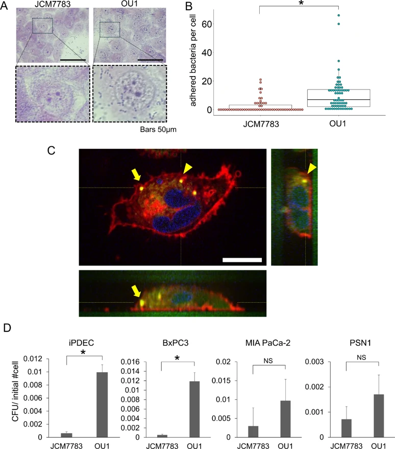
To examine the potential pathogenic role of E. faecalis in pancreas, we evaluated adhesion to immortalized normal human pancreatic ductal epithelial cells (iPDECs). Two E. faecalis strains were used to evaluate adhesion: JCM7783 and OU1. Both strains exhibited adhesion to iPDECs that persisted even after thorough washing steps, with the OU1 strain adhering to a greater extent.
Next, we examined entry of E. faecalis into iPDECs following adhesion. Image analysis confirmed that E. faecalis entered the cytoplasm of iPDECs. E. faecalis invasion of iPDECs was quantified using an invasion assay. The results demonstrated that both strains of E. faecalis entered iPDECs, with the highly adhesive OU1 strain invading to a greater extent than the JCM7783 strain. Cellular invasion by E. faecalis was also confirmed in three additional pancreatic ductal epithelial lineage cell lines: BxPC3, MIA PaCa-2, and PSN1.
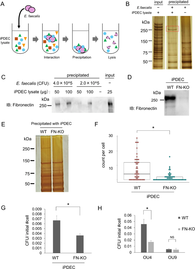
Mechanistically, bacterial expression of enterococcal fibronectin-binding protein A (EfbA) was correlated with adhesive potential of E. faecalis strains. Knockout of fibronectin 1, a binding partner of EfbA, in iPDECs resulted in suppressed E. faecalis adhesion and invasion, suggesting the importance of the EfbA-fibronectin axis in infection of pancreatic ductal epithelial lineage cells. Overall, these results suggest that E. faecalis can colonize pancreatic tissue by infecting iPDECs, at least in part, via the expression of the cell adhesion factor EfbA.
The medium used for transportation (perfusion medium) cannot be used to culture the cells again, please use a new complete medium prepared according to the cell culture conditions in the instruction manual to culture the cells.
Ask a Question
Average Rating: 5.0 | 1 Scientist has reviewed this product
Long-term usage
I have been buying Creative Bioarray products and would be happy to recommend it to others.
09 Oct 2021
Ease of use
After sales services
Value for money
Write your own review

Description: Immortalized Human Gastric Epithelial Cells-SV40 have been obtained immortalizing Human Gastric Epithelial Cells with Lenti-SV40 Lentivirus. Immortalized cells were controlled passaging side by side ...
Description: Immortalized Human Kidney Podocytes-SV40 have been obtained immortalizing Human Kidney Podocytes with Lenti-SV40 Lentivirus. Immortalized cells were controlled passaging side by side with the primary ...
Description: Nasal epithelial cells form the outermost protective layer against environmental factors. They clean, humidify, and warm inhaled air. They produces mucus, which binds particles that are subsequently ...
Description: Immortalized Human Splenic Endothelial Cells-SV40 have been obtained immortalizing Human Splenic Endothelial Cells with SV40LT expressing lentiviral particles. Immortalized cells were controlled ...
Description: Immortalized Human Corneal Epithelial Cells-SV40 have been obtained immortalizing Human Corneal Epithelial Cells with Lenti-SV40 Lentivirus. Immortalized cells were controlled passaging side by side ...
- Adipose Tissue-Derived Stem Cells
- Human Neurons
- Mouse Probe
- Whole Chromosome Painting Probes
- Hepatic Cells
- Renal Cells
- In Vitro ADME Kits
- Tissue Microarray
- Tissue Blocks
- Tissue Sections
- FFPE Cell Pellet
- Probe
- Centromere Probes
- Telomere Probes
- Satellite Enumeration Probes
- Subtelomere Specific Probes
- Bacterial Probes
- ISH/FISH Probes
- Exosome Isolation Kit
- Human Adult Stem Cells
- Mouse Stem Cells
- iPSCs
- Mouse Embryonic Stem Cells
- iPSC Differentiation Kits
- Mesenchymal Stem Cells
- Immortalized Human Cells
- Immortalized Murine Cells
- Cell Immortalization Kit
- Adipose Cells
- Cardiac Cells
- Dermal Cells
- Epidermal Cells
- Peripheral Blood Mononuclear Cells
- Umbilical Cord Cells
- Monkey Primary Cells
- Mouse Primary Cells
- Breast Tumor Cells
- Colorectal Tumor Cells
- Esophageal Tumor Cells
- Lung Tumor Cells
- Leukemia/Lymphoma/Myeloma Cells
- Ovarian Tumor Cells
- Pancreatic Tumor Cells
- Mouse Tumor Cells