

Immortalized Human Pancreatic Duct Epithelial Cells (HPDE6c7)
Cat.No.: CSC-I2298Z
Species: homo sapiens
Morphology: Polygonal
Culture Properties: Adherent
- Specification
- Background
- Scientific Data
- Q & A
- Customer Review
HPDE6c7, also designated HPDE6-E6E7c7 or H6c7, is an immortalized but non‑tumorigenic human pancreatic duct epithelial cell line established from the normal pancreatic duct of a 63‑year‑old female donor. Immortalization was achieved by transduction of a retrovirus vector carrying the E6 and E7 genes of human papillomavirus type 16 (HPV‑16) into primary normal human pancreatic duct epithelial (HPDE) cells. Single clones were isolated that retain a near‑normal genotype and phenotype of pancreatic duct epithelium.
The defining advantage of the HPDE6c7 line is its exceptionally close resemblance to normal pancreatic duct epithelium, combined with an indefinite proliferative lifespan. The cells exhibit anchorage‑dependent growth, are non‑tumorigenic in SCID mice, and express definitive ductal lineage markers including carbonic anhydrase II, MUC‑1, and cytokeratins 7, 8, 18 and 19. Importantly, HPDE6c7 cells retain normal genotypes of Ki‑ras, p53, C‑myc, and p16ᴵᴺᴷ⁴ᴬ, with the only identifiable aberration being the loss of functional p53 pathway due to E6‑mediated degradation. This near‑normal status distinguishes HPDE6c7 from pancreatic cancer cell lines, which harbor numerous driver mutations and exhibit aberrant phenotypes.
HPDE6c7 cells are a versatile and physiologically relevant model for the pancreatic ductal epithelium, widely applied in studies of pancreatic duct carcinogenesis, islet cell differentiation, and chemoprevention of pancreatic cancer. They also serve as a genetically stable baseline for stepwise introduction of PDAC‑associated alterations to model multistep pancreatic oncogenesis.
Mitochondrial Calcium Uniporter Promotes Mitophagy in Caerulein‑Treated Pancreatic Ductal Epithelial Cells
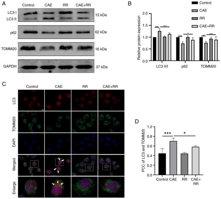
The mitochondrial calcium uniporter (MCU) is a major protein for the uptake of mitochondrial calcium to regulate intracellular energy metabolism, including processes such as mitophagy. The present study investigated the effect of the MCU on mitophagy in pancreatic ductal epithelial cells (PDECs) in acute pancreatitis (AP) in vitro. The normal human PDECs (HPDE6‑C7) were treated with caerulein (CAE) to induce AP‑like changes, with or without ruthenium red to inhibit the MCU. The mitochondrial membrane potentials (MMPs) and mitochondrial Ca2+ levels were analyzed by fluorescence. The expression levels of MCU, LC3, p62, and translocase of the outer mitochondrial membrane complex subunit 20 (TOMM20), putative kinase 1 (PINK1), and Parkin were measured by western blotting and immunofluorescence. Mitophagy was observed by confocal fluorescence microscopy and transmission electron microscopy.
The results showed that CAE increased the MCU protein expression, mitochondrial Ca2+ levels, MMP depolarization and the protein expression of mitophagy markers including the LC3II/I ratio, PINK1, and Parkin. CAE decreased the protein expression of p62 and TOMM20, and promoted the formation of mitophagosomes in HPDE6‑C7 cells. Notably, changes in these markers were reversed by inhibiting the MCU.
Ask a Question
Write your own review

Description: Immortalized Human Gastric Epithelial Cells-SV40 have been obtained immortalizing Human Gastric Epithelial Cells with Lenti-SV40 Lentivirus. Immortalized cells were controlled passaging side by side ...
Description: Immortalized Human Kidney Podocytes-SV40 have been obtained immortalizing Human Kidney Podocytes with Lenti-SV40 Lentivirus. Immortalized cells were controlled passaging side by side with the primary ...
Description: Nasal epithelial cells form the outermost protective layer against environmental factors. They clean, humidify, and warm inhaled air. They produces mucus, which binds particles that are subsequently ...
Description: Immortalized Human Splenic Endothelial Cells-SV40 have been obtained immortalizing Human Splenic Endothelial Cells with SV40LT expressing lentiviral particles. Immortalized cells were controlled ...

Description: Immortalized Human Corneal Epithelial Cells-SV40 have been obtained immortalizing Human Corneal Epithelial Cells with Lenti-SV40 Lentivirus. Immortalized cells were controlled passaging side by side ...
- Adipose Tissue-Derived Stem Cells
- Human Neurons
- Mouse Probe
- Whole Chromosome Painting Probes
- Hepatic Cells
- Renal Cells
- In Vitro ADME Kits
- Tissue Microarray
- Tissue Blocks
- Tissue Sections
- FFPE Cell Pellet
- Probe
- Centromere Probes
- Telomere Probes
- Satellite Enumeration Probes
- Subtelomere Specific Probes
- Bacterial Probes
- ISH/FISH Probes
- Exosome Isolation Kit
- Human Adult Stem Cells
- Mouse Stem Cells
- iPSCs
- Mouse Embryonic Stem Cells
- iPSC Differentiation Kits
- Mesenchymal Stem Cells
- Immortalized Human Cells
- Immortalized Murine Cells
- Cell Immortalization Kit
- Adipose Cells
- Cardiac Cells
- Dermal Cells
- Epidermal Cells
- Peripheral Blood Mononuclear Cells
- Umbilical Cord Cells
- Monkey Primary Cells
- Mouse Primary Cells
- Breast Tumor Cells
- Colorectal Tumor Cells
- Esophageal Tumor Cells
- Lung Tumor Cells
- Leukemia/Lymphoma/Myeloma Cells
- Ovarian Tumor Cells
- Pancreatic Tumor Cells
- Mouse Tumor Cells