

Immortalized Human Liver Epithelial Cells (THLE-2)
Cat.No.: CSC-I2413Z
Culture Properties: Adherent
- Specification
- Background
- Scientific Data
- Q & A
- Customer Review
DNA Profile (STR):
Amelogenin: X,Y
CSF1PO: 11,13
D13S317: 8,12
D16S539: 11,13
D5S818: 11,13
D7S820: 10,12
THO1: 7,9.3
TPOX: 8,11
vWA: 16,17
Note: Never can cells be kept at -20°C.
The THLE-2 (Transformed Human Liver Epithelial-2) cell line is an immortalized human liver epithelial cell line derived from normal adult liver tissue obtained from the left lobe of a 64-year-old male donor with no clinical evidence of cancer. The cells were immortalized by introducing the simian virus 40 (SV40) large T antigen gene via retroviral transduction, a method that markedly extends the replicative lifespan of primary hepatocytes.
One of the most distinctive and critical advantages of THLE-2 cells is their non-tumorigenic phenotype when injected into athymic nude mice, their near-diploid karyotype, and the absence of alpha-fetoprotein (AFP) expression-key features that distinguish them from classical hepatoma cell lines like HepG2. Consequently, they represent a physiologically relevant, non-malignant human in vitro model that fills a crucial gap between primary human hepatocytes and transformed liver cancer cell lines.
The THLE-2 line retains functional cytochrome P450 (CYP) pathways, as confirmed by its ability to metabolize the procarcinogens benzo[a]pyrene, N‑nitrosodimethylamine, and aflatoxin B1 to their ultimate DNA-adducting metabolites. In addition, the cells maintain several Phase II enzymes and antioxidant systems, including glutathione S‑transferases, epoxide hydrolase, superoxide dismutase and catalase, enabling investigations into both xenobiotic metabolism and cellular defense mechanisms. The characteristically low background expression of P450 genes in untransfected THLE-2 cells additionally serves as an ideal platform for stable or transient transfection of individual CYP isoforms, facilitating targeted investigations of metabolite‑mediated hepatotoxicity with minimal interference from endogenous enzyme activity.
Quantum Dots Affect Actin Cytoskeleton Reorganization, Resulting in Impaired HeLa and THLE-2 Cell Motility
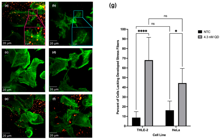
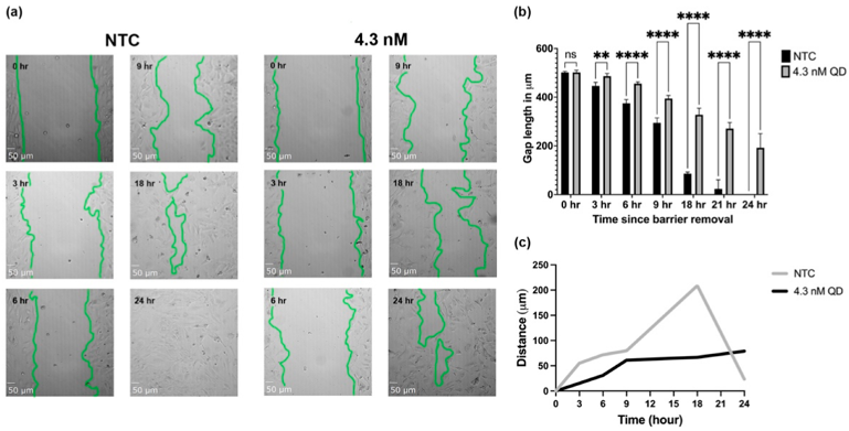
Quantum dots (QDs) are nanoparticles with intrinsic fluorescence. Recent studies have found that metal-based QDs often impart toxic effects on the biological systems they interact with. Their undefined limitations have offset their potential for biomedical application. Our study aimed to address the research gap regarding QDs' impacts on the intracellular actin cytoskeleton and the associated structures. Our XTT viability assays revealed that QDs only reduced viability in transformed human liver epithelial (THLE-2) cells, whereas HeLa cells remained viable after QD treatment. We also used confocal microscopy to evaluate the morphological changes in THLE-2 induced by QDs. We further investigated cell protrusion morphology using phalloidin-Alexa488 which selectively labels F-actin. The fluorescent microscopy of this phalloidin label revealed that QD treatment resulted in the redistribution of actin filaments within both THLE-2 and HeLa cells. We also report that the average number of focal adhesions decreased in QD-treated cells. As actin filaments at the cell are peripherally linked to the extracellular matrix via talin and integrin and are thus a crucial component of cell motility, we conducted a migration assay. The migration assay revealed that cell motility was significantly reduced in both THLE-2 and HeLa cells following QD treatment. Our findings establish that the internalization of QDs reduces cell motility by rearranging actin filaments.
Ask a Question
Write your own review
Description: Immortalized Human Gastric Epithelial Cells-SV40 have been obtained immortalizing Human Gastric Epithelial Cells with Lenti-SV40 Lentivirus. Immortalized cells were controlled passaging side by side ...

Description: Immortalized Human Kidney Podocytes-SV40 have been obtained immortalizing Human Kidney Podocytes with Lenti-SV40 Lentivirus. Immortalized cells were controlled passaging side by side with the primary ...
Description: Nasal epithelial cells form the outermost protective layer against environmental factors. They clean, humidify, and warm inhaled air. They produces mucus, which binds particles that are subsequently ...
Description: Immortalized Human Splenic Endothelial Cells-SV40 have been obtained immortalizing Human Splenic Endothelial Cells with SV40LT expressing lentiviral particles. Immortalized cells were controlled ...
Description: Immortalized Human Corneal Epithelial Cells-SV40 have been obtained immortalizing Human Corneal Epithelial Cells with Lenti-SV40 Lentivirus. Immortalized cells were controlled passaging side by side ...
- Adipose Tissue-Derived Stem Cells
- Human Neurons
- Mouse Probe
- Whole Chromosome Painting Probes
- Hepatic Cells
- Renal Cells
- In Vitro ADME Kits
- Tissue Microarray
- Tissue Blocks
- Tissue Sections
- FFPE Cell Pellet
- Probe
- Centromere Probes
- Telomere Probes
- Satellite Enumeration Probes
- Subtelomere Specific Probes
- Bacterial Probes
- ISH/FISH Probes
- Exosome Isolation Kit
- Human Adult Stem Cells
- Mouse Stem Cells
- iPSCs
- Mouse Embryonic Stem Cells
- iPSC Differentiation Kits
- Mesenchymal Stem Cells
- Immortalized Human Cells
- Immortalized Murine Cells
- Cell Immortalization Kit
- Adipose Cells
- Cardiac Cells
- Dermal Cells
- Epidermal Cells
- Peripheral Blood Mononuclear Cells
- Umbilical Cord Cells
- Monkey Primary Cells
- Mouse Primary Cells
- Breast Tumor Cells
- Colorectal Tumor Cells
- Esophageal Tumor Cells
- Lung Tumor Cells
- Leukemia/Lymphoma/Myeloma Cells
- Ovarian Tumor Cells
- Pancreatic Tumor Cells
- Mouse Tumor Cells