

SK-MEL-30
Cat.No.: CSC-C0302
Species: Homo sapiens (Human)
Morphology: adherent, epithelial-like cells growing as monolayers
Culture Properties: monolayer
- Specification
- Scientific Data
- Q & A
- Customer Review
Immunology: cytokeratin -, desmin -, endothel -, GFAP -, HMB-45 +, neurofilament -, vimentin +
Viruses: ELISA: reverse transcriptase negative; PCR: EBV -, HBV -, HCV -, HHV-8
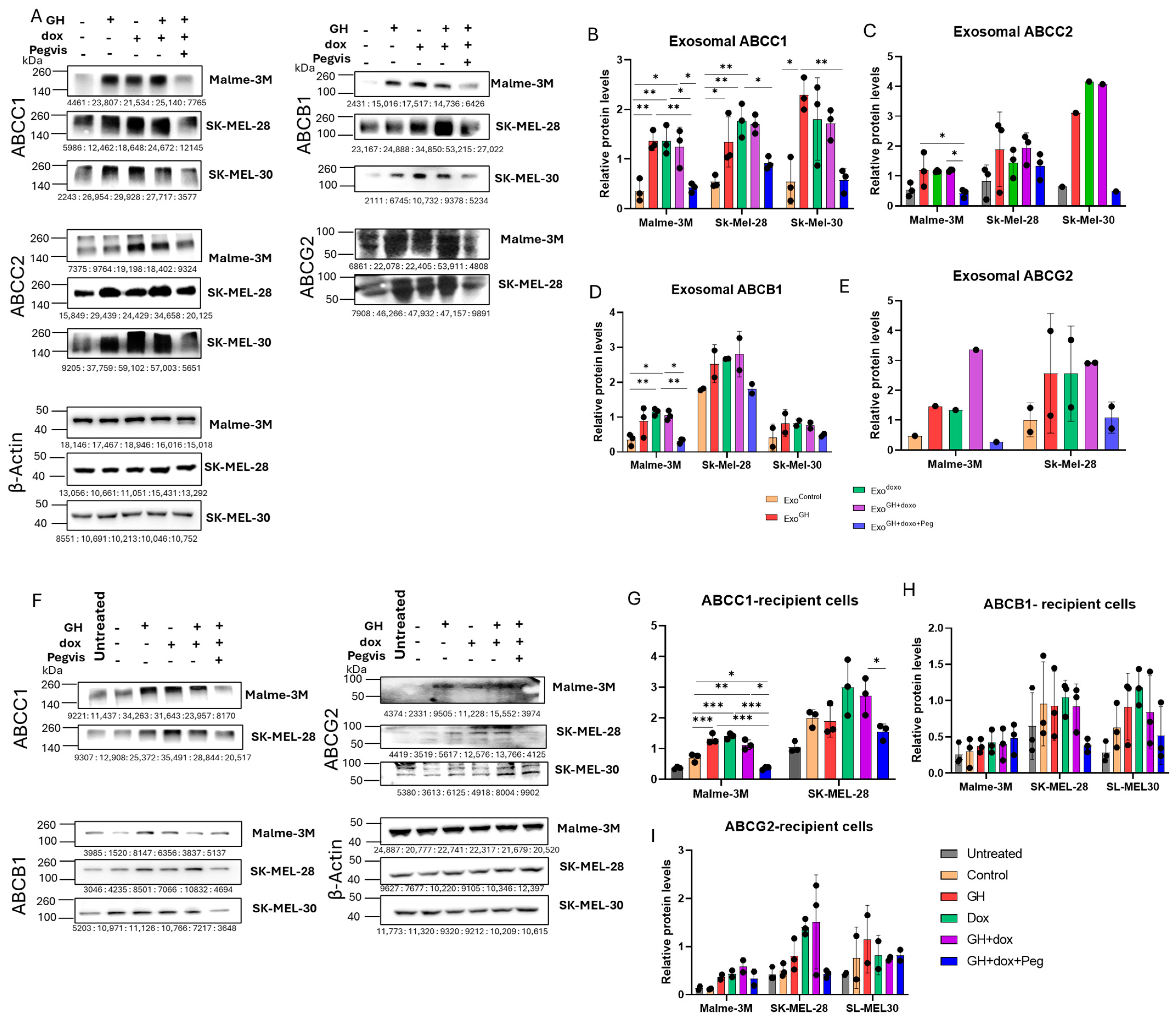
Growth Hormone Upregulates Melanoma Drug Resistance and Migration via Melanoma-Derived Exosomes
Background
This study investigates how growth hormone (GH) modulates melanoma-derived exosomes to promote drug resistance and migration. Melanoma cells were treated with GH, doxorubicin, and the GHR antagonist pegvisomant. GH-treated cells released exosomes enriched with ABC transporters ABCC1 and ABCB1, N-cadherin, and MMP2, which enhanced drug resistance and migration in recipient melanoma cells. Blocking GHR with pegvisomant reduced these exosomal cargoes, restored drug sensitivity, and attenuated migration. The findings reveal a novel mechanism by which GH drives chemoresistance and metastasis via exosomes and suggest GHR antagonism as a potential therapeutic strategy.
Key Findings
The study demonstrates that growth hormone (GH) promotes chemoresistance and migration in melanoma by altering exosomal cargo. GH treatment increased exosomal levels of ABC transporters ABCC1 and ABCB1, N-cadherin, and MMP2. Exosomes from GH-treated melanoma cells reduced drug retention and increased doxorubicin IC50 in recipient cells, indicating enhanced drug resistance. They also promoted recipient cell migration. The GHR antagonist pegvisomant reversed these effects: exosomes from cells treated with GH, doxorubicin, and pegvisomant had lower levels of these proteins, restored drug sensitivity, and attenuated migration. Blocking autocrine/paracrine GH also suppressed doxorubicin-induced exosomal ABC transporters. These findings identify a novel role for GH in packaging drug efflux pumps and EMT markers into exosomes, and suggest GHR antagonism as a strategy to overcome exosome-mediated chemoresistance in melanoma.
Contribution
Creative Bioarray contributed the SK-MEL-30 human melanoma cell line (Shirley, NY, USA) used in this study. This cell line was essential for exosome isolation, drug retention, chemosensitivity, migration assays, and western blot analyses, enabling the investigation of GH-mediated exosomal cargo and drug resistance across multiple melanoma models.
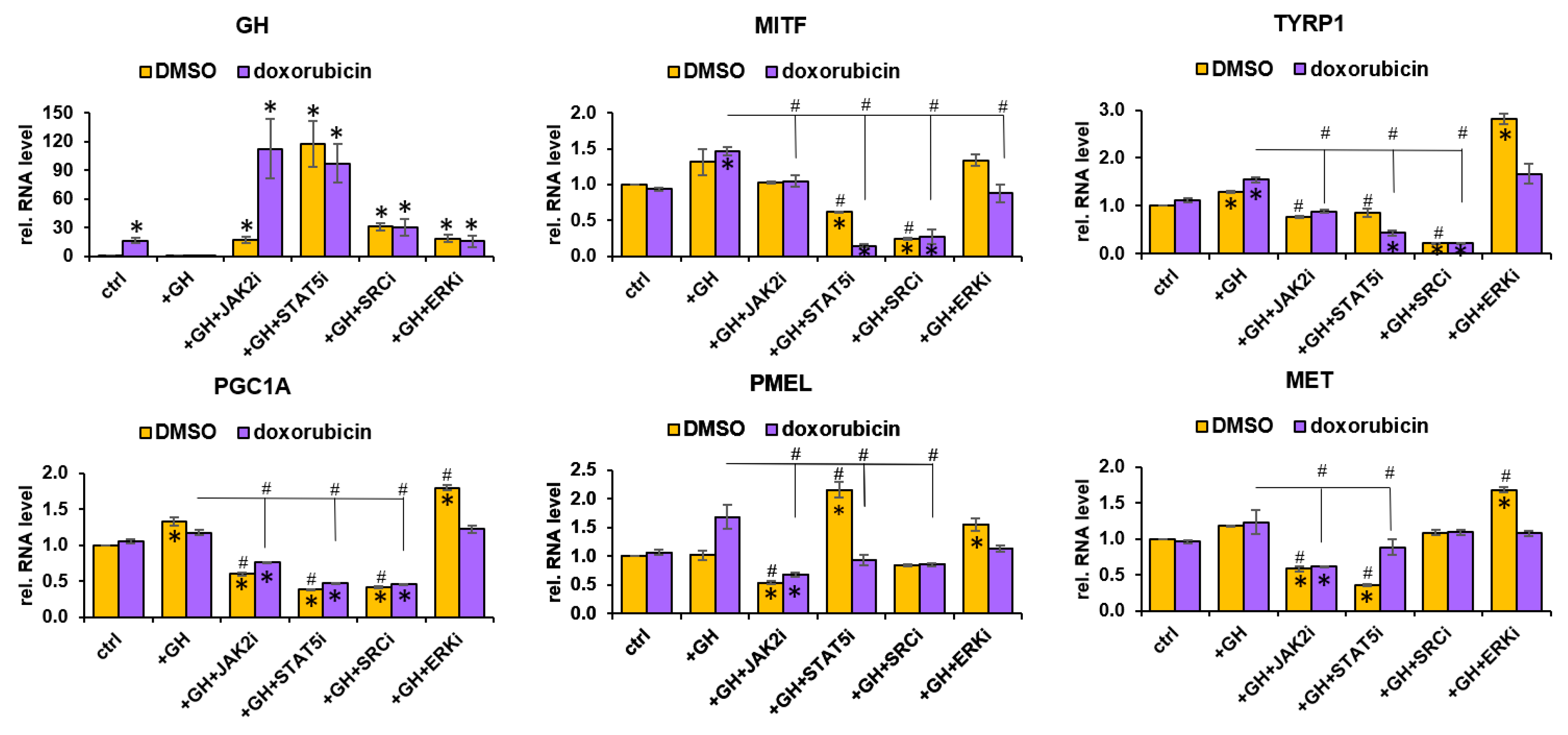
Growth Hormone Upregulates Melanocyte-Inducing Transcription Factor Expression and Activity via JAK2-STAT5 and SRC Signaling in GH Receptor-Positive Human Melanoma
Background
This study investigates how growth hormone (GH) promotes chemoresistance in GH receptor-positive human melanoma by upregulating melanocyte-inducing transcription factor (MITF). Using multiple melanoma cell lines, mouse xenografts, and public cancer datasets, the authors show that anticancer drugs increase GH/GHR and MITF expression. GH directly elevates MITF and MITF target genes, enhances melanogenesis and tyrosinase activity, and supports drug clearance pathways. Inhibitor analyses identify JAK2-STAT5 and SRC signaling as key mediators. The findings implicate the GH–GHR axis in melanoma therapy resistance and suggest GHR antagonism as a potential combinatorial strategy.
Key Findings
GH treatment significantly upregulated MITF and MITF target genes in human melanoma cells following chemotherapy, BRAF inhibitors, and tyrosine kinase inhibitors. GH also increased MITF-regulated melanogenesis and tyrosinase activity in multiple cell lines. In B16F10 mouse melanoma xenografts, bovine GH transgenic mice showed elevated MITF and target gene expression versus wild-type littermates. Pathway inhibitor analysis showed that JAK2-STAT5 and SRC activities were critical for GH-induced MITF regulation, with MAPK/ERK involvement under chemotherapy. GHR knockdown suppressed MITF, MITF targets, melanogenesis, and tyrosinase activity, sensitizing cells to drug effects. Bioinformatics analyses across GTEx, NCI60, CCLE, and TCGA datasets corroborated coexpression and correlation of GHR, MITF, and MITF targets. The study provides in vitro, in vivo, and in silico evidence that the GH–GHR axis drives MITF-regulated and ABC-transporter-mediated drug clearance, contributing to melanoma chemoresistance.
Contribution
Creative Bioarray provided the SK-MEL-30 human melanoma cell line used in the in vitro experiments. This cell line enabled the authors to test GH effects on MITF, melanogenesis, tyrosinase activity, and signaling in a BRAF wild-type melanoma context.
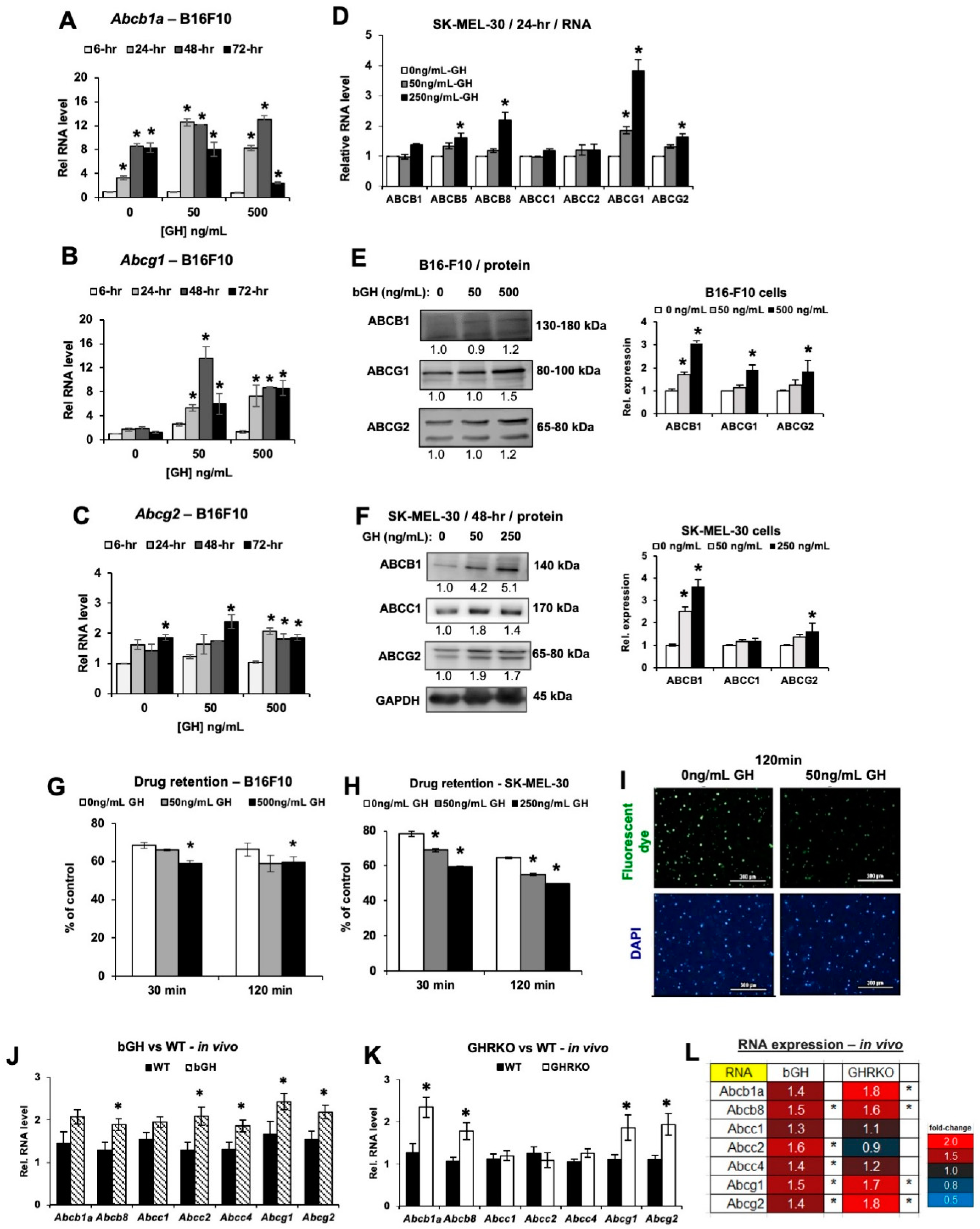
Growth Hormone Upregulates Mediators of Melanoma Drug Efflux and Epithelial-to-Mesenchymal Transition In Vitro and In Vivo
Background
This study investigates the effects of growth hormone (GH) and insulin-like growth factor-1 (IGF-1) on melanoma progression and therapy resistance. Using mouse B16-F10 and human SK-MEL-30 melanoma cells in vitro, plus syngeneic B16-F10 tumors in bovine GH transgenic (bGH) and GH receptor knockout (GHRKO) mice in vivo, the authors examined tumor growth, multidrug efflux pumps, and epithelial-to-mesenchymal transition (EMT) markers. GH treatment enhanced melanoma cell growth, drug retention, and invasion in vitro, while elevated GH upregulated drug efflux pumps and EMT markers independent of IGF-1 in vivo, revealing GH-driven intrinsic therapy resistance in melanoma.
Key Findings
GH enhanced mouse and human melanoma cell growth, drug retention, and invasion in vitro. In syngeneic models, elevated GH did not accelerate tumor size but significantly upregulated multiple ATP-binding cassette (ABC) drug-efflux pumps. GH specifically elevated Abcb1a, Abcb8, Abcg1, and Abcg2 transporters in an IGF-1-independent manner, whereas IGF-1 upregulated Abcc-type transporters, suggesting distinct and overlapping roles. Additionally, epithelial-to-mesenchymal transition (EMT) transcription markers (Zeb1, Snai1) were significantly upregulated in vivo with Cdh1 suppression, and both GH and IGF-1 proved potent EMT inducers. Combined with GHR knockdown reducing cell growth, these results provide the first in vivo validation that GH drives an intrinsic drug-resistance and metastatic phenotype in melanoma, supporting GHR antagonism as a potential therapeutic strategy alongside chemotherapy.
Contribution
Creative Bioarray provided the SK-MEL-30 human melanoma cell line (CSC-C0302) used in this study. These cells served as the key human melanoma model for assessing GH-induced signaling, ABC drug-efflux pump expression, EMT markers, proliferation, and invasion in vitro, complementing the mouse B16-F10 cells.
Ask a Question
Write your own review
- You May Also Need
Description: Established from the primary tumor (right cervical) of a 64-year-old woman with cutaneous melanoma
Description: Species: human, Caucasian male adult; Tissue: skin; Tumor: melanoma
Description: Established from the lymph node metastasis of a malignant melanoma from a 42-year-old Caucasian woman

Description: Species: human male 66 years old; Tissue: lymph node, metastasis; Tumor: melanoma
Description: Species: human - male, 31 years old, CaucasianIsoenzyme: G6PD, BProduction: melaninHistopathology: melanoma
- Adipose Tissue-Derived Stem Cells
- Human Neurons
- Mouse Probe
- Whole Chromosome Painting Probes
- Hepatic Cells
- Renal Cells
- In Vitro ADME Kits
- Tissue Microarray
- Tissue Blocks
- Tissue Sections
- FFPE Cell Pellet
- Probe
- Centromere Probes
- Telomere Probes
- Satellite Enumeration Probes
- Subtelomere Specific Probes
- Bacterial Probes
- ISH/FISH Probes
- Exosome Isolation Kit
- Human Adult Stem Cells
- Mouse Stem Cells
- iPSCs
- Mouse Embryonic Stem Cells
- iPSC Differentiation Kits
- Mesenchymal Stem Cells
- Immortalized Human Cells
- Immortalized Murine Cells
- Cell Immortalization Kit
- Adipose Cells
- Cardiac Cells
- Dermal Cells
- Epidermal Cells
- Peripheral Blood Mononuclear Cells
- Umbilical Cord Cells
- Monkey Primary Cells
- Mouse Primary Cells
- Breast Tumor Cells
- Colorectal Tumor Cells
- Esophageal Tumor Cells
- Lung Tumor Cells
- Leukemia/Lymphoma/Myeloma Cells
- Ovarian Tumor Cells
- Pancreatic Tumor Cells
- Mouse Tumor Cells