

ND7/23
Cat.No.: CSC-C9374J
Species: Mus musculus (Mouse) x Rattus norvegicus (Rat)
- Specification
- Background
- Scientific Data
- Q & A
- Customer Review
ND7/23 is a continuous hybrid cell line generated by somatic fusion of mouse neuroblastoma N18TG2 with rat dorsal root ganglion (DRG) neurons. This hybrid origin confers a sensory neuron-like phenotype while retaining the proliferative capacity and handling convenience of an immortalized line. ND7/23 cells express neuronal markers such as βIII-tubulin, neurofilament, and synaptophysin, and can be differentiated by serum withdrawal or dibutyryl cAMP to extend neurites and upregulate neuronal properties. They possess functional voltage-gated sodium and calcium channels and respond to various neuroactive compounds, making them useful for electrophysiology, calcium imaging, and neuropharmacology.
Key advantages:
- Continuous, homogenous, and scalable source; reduces batch-to-batch variability and animal use compared with primary DRG neurons.
- Retains DRG-like sensory neuron characteristics, including excitability and nociceptive signaling pathways.
- Amenable to transfection, siRNA, CRISPR, patch-clamp, high-content imaging, and high-throughput screening.
- Can be differentiated to a more mature neuronal phenotype, enabling studies of neurite outgrowth, ion channel function, and neurotoxicity.
Applications: pain research, neurotoxicity testing, ion channel pharmacology, drug discovery, and mechanistic studies of sensory neuron signaling. ND7/23 cells are also used as hosts for expressing recombinant channels or receptors, providing a controlled background for functional assays.
The Effects of Co-Culturing ND7/23 Sensory Neuron-like Cells and IFRS1 Schwann Cells on Myelination
Co-culture models of neurons and Schwann cells have been used to explore the mechanisms of myelination during development, axonal regeneration after injury, and the pathogenesis of various demyelinating neuropathies. In this study, we aimed to establish a stable co-culture system using IFRS1 cells and ND7/23 sensory neuron-like cells.
ND7/23 cells were seeded at a low density (2 × 10³/cm2) and maintained for 7 days in serum-containing medium supplemented with NGF (10 ng/mL) and the Rho kinase inhibitor Y27632 (5 μM) to promote neurite elongation. The cells were then treated with the anti-mitotic agent mitomycin C (1 μg/mL) for 12–16 h to suppress proliferative activity. Following this, the cells were co-cultured with IFRS1 cells (2 × 10⁴/cm2) and maintained at 37 °C in serum-containing medium supplemented with ascorbic acid (50 μg/mL), NGF (10 ng/mL), and ciliary neurotrophic factor (10 ng/mL).
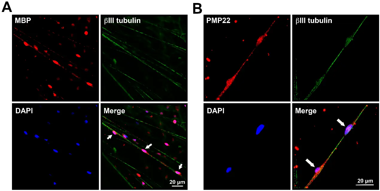
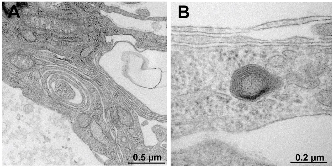
Double-immunofluorescence staining performed on day 21 of the co-culture revealed myelin protein 22- or myelin basic protein-immunoreactive IFRS1 cells surrounding βIII tubulin-immunoreactive neurites emerging from ND7/23 cells. Myelin formation was further confirmed via Sudan Black B staining and electron microscopy.
Ask a Question
Write your own review
Description: Hybridoma producing a monoclonal antibody against tetrodotoxin.
Description: MoAb to Clostridium perfringens alpha-toxin (phospholipase C).
Description: Strain: BALB/cJ miceProduction: Monoclonal antibody(IgG2b), reactive to human granulocytes, monocytes and null cells, is produced.Histopathology: hybridoma; mouse(myeoloma)Note: Immunized with human ...
Description: Hybridoma producing a monoclonal antibody against gonyautoxin.
Description: Cells are a subclone derived from B13.29 (first described as clone 9.9) by fusion of BALB/c spleen cells with SP2/0-AG14 myeloma cells; this hybridoma is a non-producer. B9 is a mouse B cell ...
- Adipose Tissue-Derived Stem Cells
- Human Neurons
- Mouse Probe
- Whole Chromosome Painting Probes
- Hepatic Cells
- Renal Cells
- In Vitro ADME Kits
- Tissue Microarray
- Tissue Blocks
- Tissue Sections
- FFPE Cell Pellet
- Probe
- Centromere Probes
- Telomere Probes
- Satellite Enumeration Probes
- Subtelomere Specific Probes
- Bacterial Probes
- ISH/FISH Probes
- Exosome Isolation Kit
- Human Adult Stem Cells
- Mouse Stem Cells
- iPSCs
- Mouse Embryonic Stem Cells
- iPSC Differentiation Kits
- Mesenchymal Stem Cells
- Immortalized Human Cells
- Immortalized Murine Cells
- Cell Immortalization Kit
- Adipose Cells
- Cardiac Cells
- Dermal Cells
- Epidermal Cells
- Peripheral Blood Mononuclear Cells
- Umbilical Cord Cells
- Monkey Primary Cells
- Mouse Primary Cells
- Breast Tumor Cells
- Colorectal Tumor Cells
- Esophageal Tumor Cells
- Lung Tumor Cells
- Leukemia/Lymphoma/Myeloma Cells
- Ovarian Tumor Cells
- Pancreatic Tumor Cells
- Mouse Tumor Cells