
Mouse Embryonic Fibroblasts
Cat.No.: CSC-C8043L
Species: Mouse
Source: Embryo
Cell Type: Fibroblast
- Specification
- Background
- Scientific Data
- Q & A
- Customer Review
Mouse embryonic fibroblasts (MEFs) are fibroblasts isolated from mouse embryos. They can be isolated at the early stages of mouse embryonic development (day 9–14 gestation period). It's a specific kind of fibroblast that differentiated from mesenchymal cells during the embryogenesis. Morphologically, they have spindle or stellate shapes with large oval-shaped nuclei and prominent nucleoli. MEFs are widely used in a variety of research applications. They are used as feeder cells to support ESCs or iPSCs to remain undifferentiated and support their long-term culture and characterization. In addition, MEFs can be used for functional studies of genes, signaling, inflammation, immunity and disease models (fibrosis, cancer, etc.).
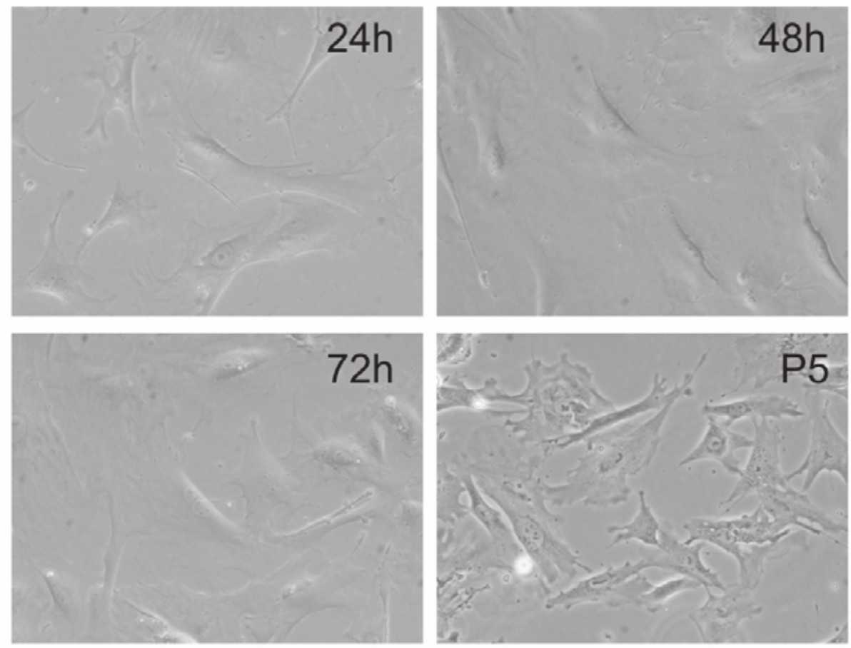 Fig. 1. Morphology of primary mouse embryonic fibroblasts (MEFs) (Huang E, Bi Y, et al., 2012).
MEFs Undergo Reversible AMPylation of the ER Chaperon BiP
The endoplasmic reticulum (ER) relies on the chaperone BiP to regulate protein folding and stress responses. FicD, a bifunctional enzyme, modulates BiP activity via AMPylation/deAMPylation, but its role in cellular stress adaptation remains unclear. Gluen et al. investigated how FicD influences mouse embryonic fibroblasts (MEFs) during pharmacologically and metabolically induced ER stress, particularly in glucose fluctuations.
To analyze BiP AMPylation, they produced immortalized FLAG-tagged FicDF/F and FicD−/− MEFs. The FicD protein was not detectable by Western blot analysis for endogenous expression. However, using RT-qPCR, they could confirm that FicDF/F MEFs expressed a functional FicD that can mediate reversible BiP AMPylation, which is absent in FicD−/− cells (Fig. 1B). Addition of CHX, a translation inhibitor, induced a higher level of BiP AMPylation in FicDF/F compared to FicD−/− MEFs. However, treatment of FicDF/F MEFs with DTT, a reducing agent, led to the complete reversal of BiP AMPylation after 60 min in a stress-dependent manner (Fig. 1B). In a time course, BiP AMPylation was no longer detectable in both stressed groups (TG irreversible stress and DTT reversible stress), but reappeared only after glucose recovery in DTT-treated cells until 8 h (Fig. 1C, D). They also detected increased BiP AMPylation in FicDF/F MEFs after glucose starvation, a physiological stress condition, which returned to basal levels when glucose was replenished (Fig. 1E).
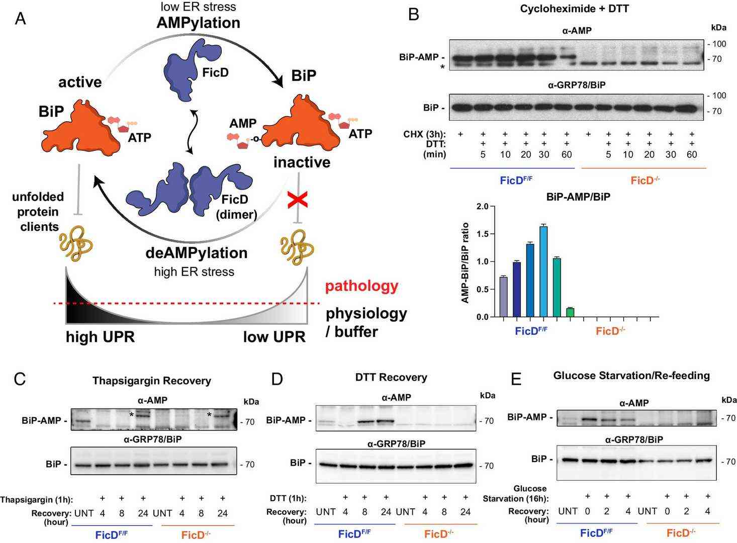 Fig. 1. AMPylation of BiP and response to ER stress in MEFs (Gluen B, Bievins A, et al., 2024).
Berberine Ameliorates Mitochondrial Dysfunction in a PINK1 Knockout Model
Mitophagy is important for maintaining cellular homeostasis and is involved in the pathogenesis of neurodegenerative diseases such as Parkinson's disease. Berberine is a natural alkaloid that may play a role in regulating mitochondrial function. Um et al. investigated whether berberine can induce mitophagy via AMPK activation and rescue mitochondrial dysfunction in PINK1 knockout cells, offering a potential therapy for neurodegenerative diseases.
Mitophagy can improve mitochondrial function in different model of mitochondrial dysfunction. To see whether berberine can affect mitochondrial function in the same way, they treated PINK1 knockout (PINK1 KO) MEFs, which display a decreased mitochondrial membrane potential, increased ROS and respiration, with berberine. After 24 h, they measured mitophagy by SDHB levels. In PINK1 KO MEFs, SDHB was decreased at 40 μM berberine, and there was a significant decrease at 80 μM (Fig. 2A). PINK1 KO MEFs showed a 46% reduction in mitochondrial membrane potential as expected (Fig. 2B). Treatment with berberine restored potential, reduced ROS to wild-type levels (Fig. 2C), and rescued mitochondrial respiration (Fig. 2D). Basal respiration and maximal respiration were increased by 50% and 113%, respectively (Fig. 2E and F). This suggests that berberine reversed mitochondrial dysfunction in PINK1 KO MEFs. They wanted to see if this effect was mediated by AMPK, so they used the AMPK inhibitor compound C. Restoration of mitochondrial potential and the decrease in ROS by berberine were abolished by compound C (Fig. 2G and H), which confirmed that berberine acts through AMPK-dependent mitophagy.
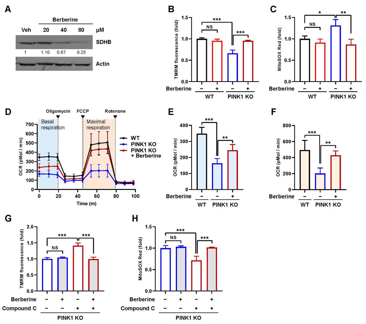 Fig. 2. Effect of berberine on mitochondrial dysfunction in PINK1 KO MEFs (Um J-H, Lee K-M, et al., 2024).
Ask a Question
Write your own review
- You May Also Need

Description: C57BL/6-GFP Mouse Skeletal Muscle Microvascular Endothelial Cells from Creative Bioarray are isolated from C57BL/6-Tg (CAG-EGFP) 1Osb/J mouse skeletal muscle tissue of pathogen-free laboratory mice. ...

Description: eNOS KO Mouse Stomach Epithelial Cells from Creative Bioarray are isolated from stomach tissue of pathogen-free laboratory mice. eNOS KO Mouse Stomach Epithelial Cells are grown in a T25 tissue ...

Description: eNOS KO Mouse Liver Fibroblasts from Creative Bioarray are isolated from liver tissue of pathogen-free laboratory mice. eNOS KO Mouse Liver Fibroblasts are grown in T75 tissue culture flasks ...
Description: C57BL/6-GFP Mouse Corneal Epithelial Cells from Creative Bioarray are isolated from C57BL/6-GFP-Tg(CAG-EGFP)1Osb/J mouse corneal tissue of pathogen-free laboratory mice. C57BL/6-GFP Mouse Corneal ...

Description: BALB/c Mouse Retinal Microvascular Endothelial Cells from Creative Bioarray are isolated from retinal tissue of pathogen-free laboratory mice. BALB/c Mouse Retinal Microvascular Endothelial Cells are ...