
LA-N-1
Cat.No.: CSC-C9417J
Species: Homo sapiens (Human)
Source: Bone Marrow Metastasis
- Specification
- Scientific Data
- Q & A
- Customer Review
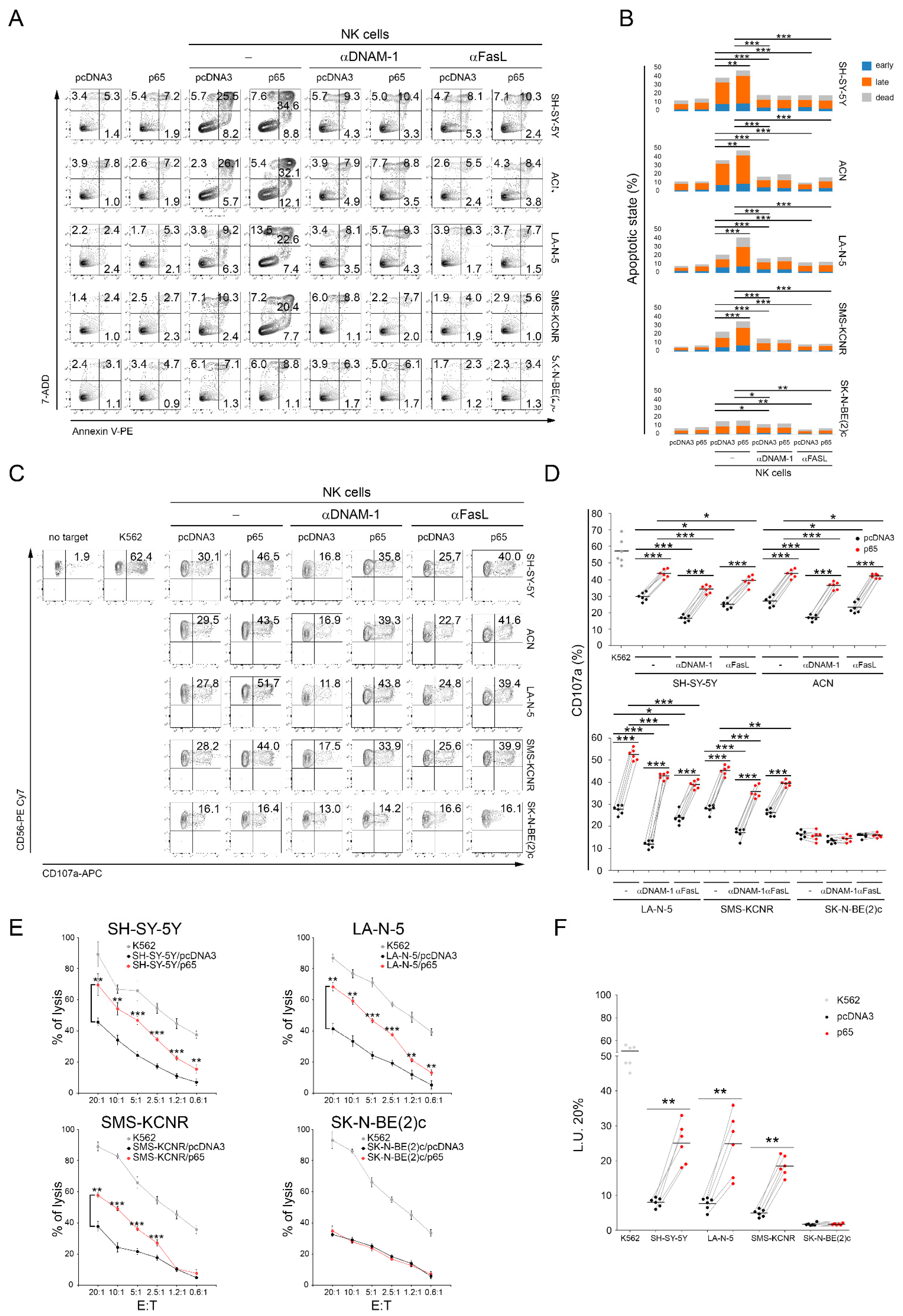
Enhancement of Neuroblastoma NK-Cell-Mediated Lysis through NF-kB p65 Subunit-Induced Expression of FAS and PVR, the Loss of Which Is Associated with Poor Patient Outcome
Background
High-risk neuroblastoma evades NK cells partly via decreased activating ligands and death receptors. This study examined whether NF-kB p65 upregulates Fas and PVR in neuroblastoma cells. Transient p65 overexpression increased surface Fas and PVR, enhancing NK-cell-mediated apoptosis, recognition, and killing; blocking DNAM-1 or FasL reduced these effects. IFNγ/TNFα also increased Fas and apoptosis but induced MHC-I and IDO1, impairing degranulation. Array CGH in patient tumors showed FAS/PVR loss correlated with poor survival independent of stage, suggesting prognostic and therapeutic relevance.
Key Findings
Transient overexpression of NF-kB p65 in neuroblastoma cell lines upregulated surface Fas and PVR, the DNAM-1 ligand, and significantly increased NK-cell-mediated apoptosis, recognition, and killing; blocking DNAM-1 or FasL reduced these effects, confirming both pathways. This effect depended on intact FAS and PVR genes, as SK-N-BE(2)c cells with both genes lost remained unaffected. IFNγ and TNFα treatment increased Fas and NK-cell-mediated apoptosis but reduced NK-cell degranulation, associated with strong MHC class I and IDO1 upregulation, indicating cytokine-induced immune evasion. High-resolution array CGH on NB patient tumors showed that loss of FAS and/or PVR correlated with poor survival independent of disease stage; 77% of patients with FAS/PVR loss died versus 19% with intact genes. The findings identify FAS/PVR status as a prognostic biomarker and suggest that transient NF-kB activation to restore Fas/PVR may enhance NK-cell immunotherapy in neuroblastoma.
Contribution
Creative Bioarray provided the LA-N-1 human neuroblastoma cell line used in this study. This cell line was included in the NB panel for array CGH and functional assays, contributing to analyses of FAS/PVR gene status, p65-mediated Fas/PVR induction, and NK-cell-mediated cytotoxicity.
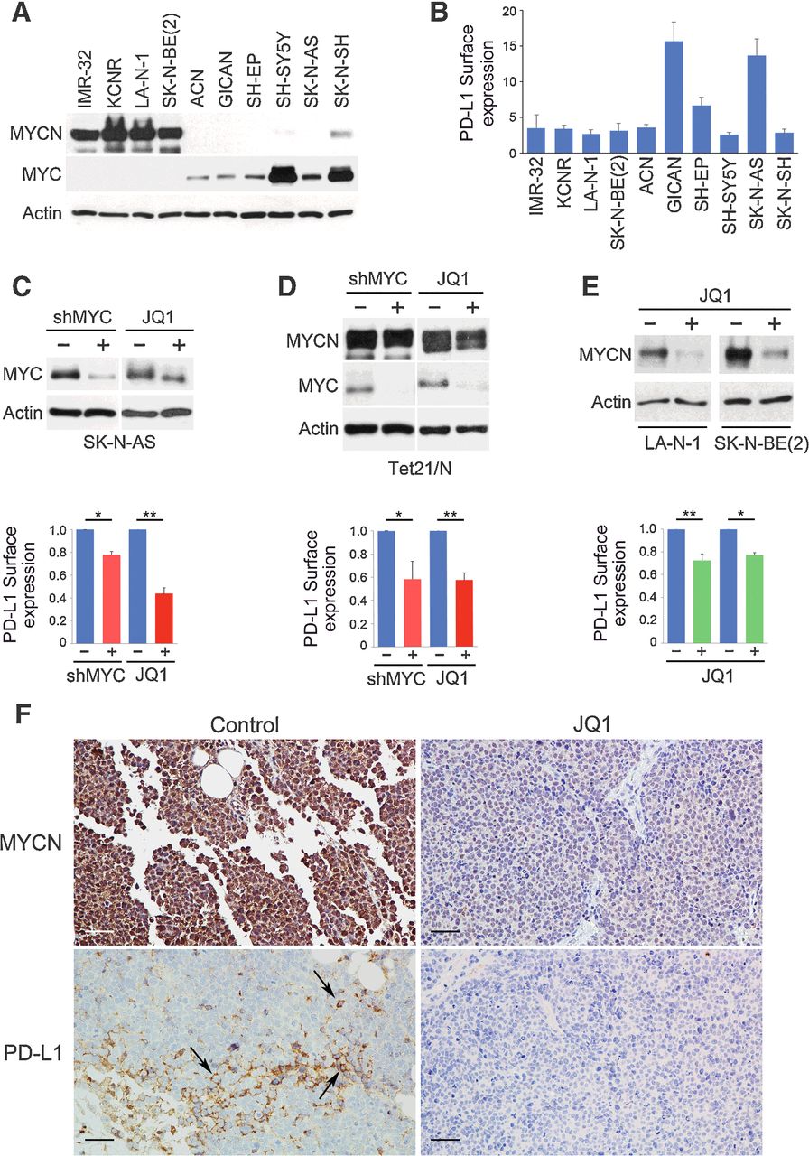
PD-L1 Is a Therapeutic Target of the Bromodomain Inhibitor JQ1 and, Combined with HLA Class I, a Promising Prognostic Biomarker in Neuroblastoma
Background
To define risk stratification and immunotherapy potential in neuroblastoma, this study evaluated PD-L1 and HLA class I on tumor cells and PD-1/LAG3 on tumor-infiltrating lymphocytes. In 77 specimens, the combination of PD-L1 and HLA class I density predicted overall survival and remained prognostic in multivariate analysis. MYC and MYCN controlled PD-L1 expression in neuroblastoma cells in vitro and in vivo; the bromodomain inhibitor JQ1 reduced MYCN/MYC and PD-L1 surface levels. These findings suggest targeting PD-L1 via MYC/MYCN inhibition may restore antitumor immunity in high-risk neuroblastoma.
Key Findings
In situ analysis of 77 neuroblastoma samples showed that PD-L1 and HLA class I tumor cell densities are inversely correlated, and their combination stratifies patients into good-prognosis (high HLA-I, low/absent PD-L1) and poor-prognosis (low HLA-I, high/absent PD-L1) groups. Multivariate Cox regression identified the PD-L1/HLA-I combination as an independent prognostic biomarker for overall survival (P = 0.0448), regardless of T-cell infiltration, MYCN amplification, INSS stage, or age. Mechanistically, MYC and MYCN regulate PD-L1 expression: MYC shRNA or JQ1 treatment reduced PD-L1 surface expression in neuroblastoma cell lines, and JQ1 decreased PD-L1 in SK-N-BE(2) xenografts. In 477 primary neuroblastomas, PD-L1 transcript correlated with MYC. Thus, pharmacologic MYC/MYCN inhibition may target PD-L1 and enhance antitumor immunity.
Contribution
Creative Bioarray provided the LA-N-1 human neuroblastoma cell line, which was used in vitro to assess MYC/MYCN-driven PD-L1 expression and response to the bromodomain inhibitor JQ1. This reagent supported the mechanistic experiments linking MYC/MYCN inhibition to reduced PD-L1, contributing to the study's therapeutic conclusions.
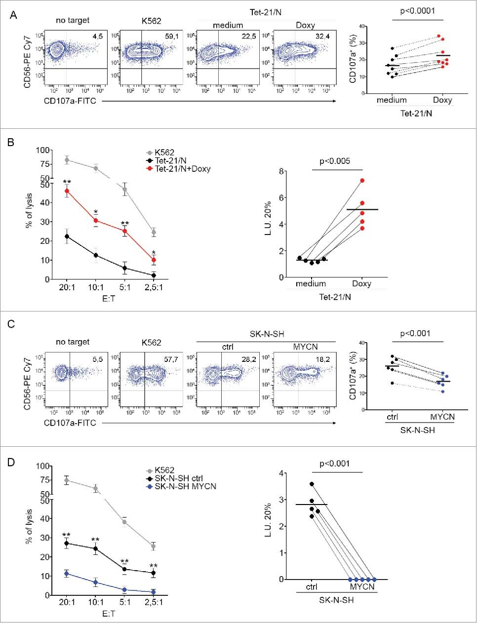
MYCN is an immunosuppressive oncogene dampening the expression of ligands for NK-cell-activating receptors in human high-risk neuroblastoma
Background
This study investigates how MYCN amplification in human high-risk neuroblastoma suppresses natural killer cell–activating ligands. In neuroblastoma cell lines and patient samples, MYCN expression inversely correlated with MICA, MICB, ULBPs and PVR. MYCN downregulation increased ligand expression and enhanced NK-cell recognition and lysis, whereas MYCN overexpression reduced these ligands and NK susceptibility. The findings identify MYCN as an immunosuppressive oncogene and suggest MYCN levels may predict the efficacy of NK-cell immunotherapy.
Key Findings
The study shows MYCN expression inversely correlates with ligands for NK-cell-activating receptors in human neuroblastoma. Across 12 neuroblastoma cell lines and 12 primary patient samples, higher MYCN levels were associated with lower surface expression of MICA, MICB, ULBP1, ULBP2/5/6, ULBP3 and PVR. In the MYCN-inducible Tet-21/N cell line, doxycycline-mediated MYCN downregulation rapidly increased these activating ligands and rendered tumor cells more susceptible to NK-cell degranulation and cytotoxicity. Conversely, transfection of MYCN into non-amplified SK-N-SH cells reduced ligand expression and decreased NK-cell recognition and lysis. MYCN expression correlated inversely with c-MYC and directly with p53/MDM2, suggesting involvement of these pathways. Primary tumors confirmed inverse MYCN–ligand correlations at mRNA and protein levels. These results define MYCN as an immunosuppressive oncogene that promotes immune escape and suggest MYCN levels as a biomarker for NK-cell immunotherapy.
Contribution
Creative Bioarray contributed by supplying the LA-N-1 human neuroblastoma cell line, which was included in the panel of NB cell lines used to analyze MYCN expression and NK-cell-activating receptor ligands. This cell line helped support the inverse correlation and functional findings.
Ask a Question
Write your own review
Description: Subline of GOTO. Protein-free medium adapted. Cell growth is slow.
Description: Established from the adrenal tumor tissue resected after treatment from a 20-month-old boy of European origin with neuroblastoma (stage III) in 1991
Description: established from the bone marrow metastasis of a 1-year-old female patient with adrenal gland neuroblastoma negative
Description: Established from the primary adrenal tumor resected prior to treatment from a 3-year-old boy with rapidly progressing Stage III neuroblastoma in 1989; described to lack MYCN (NMYC, N-myc) ...
Description: Established from a neuroblastoma metastasis at an adrenal site of a 4-year-old white boy in 1986; cells were described to express neuron-specific enolase and synaptophysin (but neither GFAP nor S-100 ...
- Adipose Tissue-Derived Stem Cells
- Human Neurons
- Mouse Probe
- Whole Chromosome Painting Probes
- Hepatic Cells
- Renal Cells
- In Vitro ADME Kits
- Tissue Microarray
- Tissue Blocks
- Tissue Sections
- FFPE Cell Pellet
- Probe
- Centromere Probes
- Telomere Probes
- Satellite Enumeration Probes
- Subtelomere Specific Probes
- Bacterial Probes
- ISH/FISH Probes
- Exosome Isolation Kit
- Human Adult Stem Cells
- Mouse Stem Cells
- iPSCs
- Mouse Embryonic Stem Cells
- iPSC Differentiation Kits
- Mesenchymal Stem Cells
- Immortalized Human Cells
- Immortalized Murine Cells
- Cell Immortalization Kit
- Adipose Cells
- Cardiac Cells
- Dermal Cells
- Epidermal Cells
- Peripheral Blood Mononuclear Cells
- Umbilical Cord Cells
- Monkey Primary Cells
- Mouse Primary Cells
- Breast Tumor Cells
- Colorectal Tumor Cells
- Esophageal Tumor Cells
- Lung Tumor Cells
- Leukemia/Lymphoma/Myeloma Cells
- Ovarian Tumor Cells
- Pancreatic Tumor Cells
- Mouse Tumor Cells