
KU-19-19
Cat.No.: CSC-C0442
Species: Homo sapiens (Human)
Source: Bladder
Morphology: epithelial-like cells growing adherently in monolayers
Culture Properties: monolayer
- Specification
- Background
- Scientific Data
- Q & A
- Customer Review
Immunology: cytokeratin +, cytokeratin-7 +, cytokeratin-8 +, cytokeratin-17 +, cytokeratin-18 +, cytokeratin-19 +, desmin -, endothel -, EpCAM -, GFAP -, neurofilament -, vim
KU-19-19 is a human bladder carcinoma cell line with distinct advantages for studying transitional cell carcinoma biology, tumor microenvironment interactions, and cytokine-mediated signaling. This epithelial cell line was established in 1993 by a Japanese research team from an invasive transitional cell carcinoma of the bladder (grade 3, pT3b) obtained from a 76-year-old male patient who exhibited marked leukocytosis. The cell line grows adherently as epithelial-like cells in monolayers under standard culture conditions, with a doubling time of approximately 48 hours.
A defining feature of KU-19-19 cells is their ability to constitutively produce and secrete multiple hematopoietic growth factors. Conditioned medium from KU-19-19 cultures contains high levels of granulocyte colony-stimulating factor (G-CSF, exceeding 5 ng/mL), as well as GM-CSF, M-CSF, stem cell factor (SCF), interleukin-6 (IL-6), and interleukin-8 (IL-8). These secreted factors vigorously stimulate proliferation of growth factor-dependent hematopoietic cell lines in a dose-dependent manner, confirming their functional biological activity. This robust secretory profile makes KU-19-19 a practical source for G-CSF purification and a versatile platform for tumor-hematopoietic cell interaction studies.
KU-19-19 cells exhibit constitutive activation of NF-κB, a transcription factor frequently dysregulated in advanced malignancies. When implanted, these cells form tumors in nude mice, associated with severe neutrophilia in peripheral blood, recapitulating the paraneoplastic leukocytosis observed in the original patient. Numerous studies have leveraged KU-19-19 cells to evaluate drug candidates such as the NF-κB inhibitor DHMEQ and natural compounds active against bladder cancer progression, demonstrating their broad utility in anticancer research.
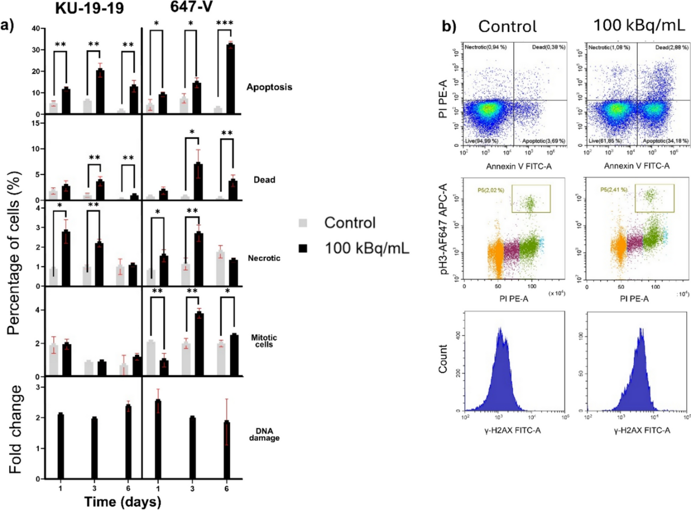
Cytotoxicity of 212Pb-Labeled Anti-PTK7 Antibody in 2D Adherent and 3D Multicellular Bladder Cancer Models
This study evaluated the preclinical efficacy of [212Pb]Pb-TCMC-chOI-1, a 212Pb-labeled antibody targeting PTK7, for targeted alpha-emitting radionuclide therapy in bladder cancer using 2D adherent cultures (clonogenic assay) and 3D multicellular spheroid models (spheroid growth inhibition).
[212Pb]Pb-TCMC-chOI-1 treatment resulted in activity- and time-dependent cytotoxicity, with enhanced sensitivity observed in cell lines with higher PTK7 levels. In clonogenic assays, the activity concentration required for 50% growth reduction was 48-74 kBq/mL, corresponding to 22-51 bound and 9-16 internalized 212Pb atoms per cell. In 3D models, similar therapeutic effects were observed despite significantly lower activities (values of approximately 1 and 30 kBq/mL for KU-19-19 and 647-V cells, respectively), suggesting a more pronounced cross-fire effect. Flow cytometry demonstrated treatment-induced DNA damage, cell cycle perturbations and cell death, with response patterns correlating with overall treatment sensitivity. RT-112 and KU-19-19 cells showed superior responses compared to 647-V and T-24 cells, consistent with their higher PTK7 expression.
These findings support PTK7 as a therapeutic target for bladder cancer and demonstrate the potential of [212Pb]Pb-TCMC-chOI-1 for targeted alpha-emitting radionuclide therapy.
![Cytotoxicity of [212Pb]Pb-TCMC-chOI-1 in 3D multicellular models.](/upload/images/ku-19-19-casestudy-1.webp)
Ask a Question
Write your own review
- You May Also Need
Description: Established from a male patient with a primary urothelial bladder carcinoma (malignant, grade 2)

Description: These cells were thought to be "stablished from the malignant urinary bladder carcinoma of a 66-year-old Caucasian man (stage pTa G3) in 1989"; same donor as for cell line BT-A; however, DNA ...
Description: Established from the tumor specimen resected from the urinary bladder transitional cell carcinoma (undifferentiated, grade IV) of a 67-year-old woman in 1974 (without therapy)

Description: Contains the unusual type A isoenzyme of glucose-6-phosphate dehydrogenase.

Description: Species: human - male, 44 years old, CaucasianTumorigenecity: yes, in mice and hamstersIsoenzyme: G6PD, BProduction: fibrinolytic activityHistopathology: carcinomaNote: the cells will grow in soft ...
Description: Species: human - female, 58 years old, CaucasianTumorigenecity: yes, in mice and hamstersIsoenzyme: G6PD,BProduction: fibrinolytic activity; interferonHistopathology: carcinoma; cancer; Grade 3Note: ...
- Adipose Tissue-Derived Stem Cells
- Human Neurons
- Mouse Probe
- Whole Chromosome Painting Probes
- Hepatic Cells
- Renal Cells
- In Vitro ADME Kits
- Tissue Microarray
- Tissue Blocks
- Tissue Sections
- FFPE Cell Pellet
- Probe
- Centromere Probes
- Telomere Probes
- Satellite Enumeration Probes
- Subtelomere Specific Probes
- Bacterial Probes
- ISH/FISH Probes
- Exosome Isolation Kit
- Human Adult Stem Cells
- Mouse Stem Cells
- iPSCs
- Mouse Embryonic Stem Cells
- iPSC Differentiation Kits
- Mesenchymal Stem Cells
- Immortalized Human Cells
- Immortalized Murine Cells
- Cell Immortalization Kit
- Adipose Cells
- Cardiac Cells
- Dermal Cells
- Epidermal Cells
- Peripheral Blood Mononuclear Cells
- Umbilical Cord Cells
- Monkey Primary Cells
- Mouse Primary Cells
- Breast Tumor Cells
- Colorectal Tumor Cells
- Esophageal Tumor Cells
- Lung Tumor Cells
- Leukemia/Lymphoma/Myeloma Cells
- Ovarian Tumor Cells
- Pancreatic Tumor Cells
- Mouse Tumor Cells