HT-1197
Cat.No.: CSC-C9438L
Species: Homo sapiens (Human)
Source: Bladder
Culture Properties: monolayer
- Specification
- Background
- Scientific Data
- Q & A
- Customer Review
Tumorigenecity: yes, in mice and hamsters
Isoenzyme: G6PD, B
Production: fibrinolytic activity
Histopathology: carcinoma
Note: the cells will grow in soft agar
vWA: 16,18
FGA: 20,23
Amelogenin: X,Y
TH01: 6,9.3
TPOX: 11,12
CSF1P0: 11,12
D5S818: 12
D13S317: 11,12
D7S820: 11,12
HT-1197 is a bladder carcinoma cell line derived from human bladder carcinoma. It was derived from a high-grade transitional cell carcinoma of urinary bladder of unknown primary tumor origin. These cells exhibit features of an invasive phenotype commonly seen in urothelial cancers and serve as an in vitro bladder cancer model to study tumor progression and response to therapies.
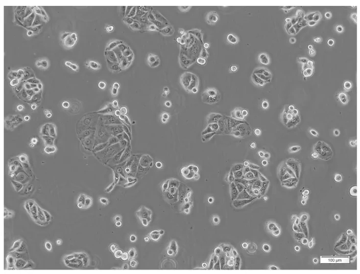
HT-1197 cells are epithelial-like adherent cells that form monolayers. Morphologically, these cells are polygonal to cobblestone, typical of bladder cancer cells. HT-1197 cells have been shown to proliferate rapidly and can be maintained with normal growth rates. Karyotyping and gene expression studies have shown HT-1197 cells to have chromosomal abnormalities and dysregulated expression of genes involved in important cellular pathways.
HT-1197 cells have been used as models to study bladder cancer cell invasion and metastasis. Studies focus on regulation of cell-cell and cell-matrix adhesion, migration, and expression of molecules involved in these pathways. These cells have also been used to study the therapeutic response of urothelial carcinoma cells to chemotherapeutic drugs, targeted therapeutics and drug combinations.
Pulsed Electromagnetic Field Therapy Alters the Genomic Profile of Bladder Cancer Cell Line HT-1197
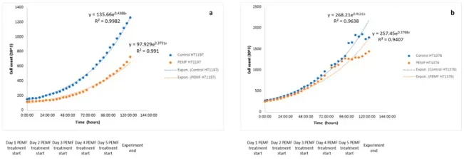
Pulsed electromagnetic field (PEMF) therapy uses magnetic waveform energy for targeted treatment and has shown promise in various cancers. Given the invasive nature of current bladder cancer treatments, Sandberg et al. investigated PEMF's effects on bladder cancer cells in vitro.
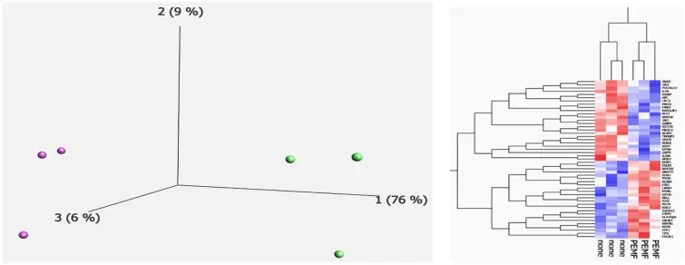
In HT-1197 cells, PEMF treatment significantly reduced proliferation compared to controls (growth rate: 0.0152 vs. 0.0176; p < 0.05, CI 4.8-27.5; R² = 0.99 for both; Fig. 1 and Fig. 2a). In contrast, HT-1376 cells showed no significant growth difference (p = 0.353, CI -108.7-39.4; R² = 0.96 and 0.94; Fig. 2b). Principal component analysis revealed complete separation between PEMF-treated and control HT-1197 cells, with PC1 accounting for 76% of variation (Fig. 3a). Transcriptome analysis identified 49 differentially expressed transcripts, visualized by heatmap showing distinct up- and downregulation patterns in PEMF-treated cells (Fig. 3b).
Ask a Question
Write your own review
- You May Also Need

Description: Contains the unusual type A isoenzyme of glucose-6-phosphate dehydrogenase.
Description: Species: human - female, 58 years old, CaucasianTumorigenecity: yes, in mice and hamstersIsoenzyme: G6PD,BProduction: fibrinolytic activity; interferonHistopathology: carcinoma; cancer; Grade 3Note: ...

Description: Established from the bladder tumor of an 84-year-old Caucasian woman following transurethral tumor resection in 1977
Description: Species: human - male, 58 years old, Caucasian, SwedishTumorigenecity: noIsoenzyme: Me-2, 1-2; PGM3, 2; PGM1, 1; ES-D, 1; AK-1, 1; G6PD, B; GLO-1Histopathology: carcinoma, transitional cell

Description: Derived from malignant ascitic fluid of a 75-year-old man with urinary bladder carcinoma in 1988; described as being of epithelial origin with morphologically distinct cells (polygonal to ...
Description: Derived from the invasive solid transitional cell carcinoma of the bladder of a 82-year-old Caucasian woman (grade IV, stage C); cell growth described as being inhibited by EGF
- Adipose Tissue-Derived Stem Cells
- Human Neurons
- Mouse Probe
- Whole Chromosome Painting Probes
- Hepatic Cells
- Renal Cells
- In Vitro ADME Kits
- Tissue Microarray
- Tissue Blocks
- Tissue Sections
- FFPE Cell Pellet
- Probe
- Centromere Probes
- Telomere Probes
- Satellite Enumeration Probes
- Subtelomere Specific Probes
- Bacterial Probes
- ISH/FISH Probes
- Exosome Isolation Kit
- Human Adult Stem Cells
- Mouse Stem Cells
- iPSCs
- Mouse Embryonic Stem Cells
- iPSC Differentiation Kits
- Mesenchymal Stem Cells
- Immortalized Human Cells
- Immortalized Murine Cells
- Cell Immortalization Kit
- Adipose Cells
- Cardiac Cells
- Dermal Cells
- Epidermal Cells
- Peripheral Blood Mononuclear Cells
- Umbilical Cord Cells
- Monkey Primary Cells
- Mouse Primary Cells
- Breast Tumor Cells
- Colorectal Tumor Cells
- Esophageal Tumor Cells
- Lung Tumor Cells
- Leukemia/Lymphoma/Myeloma Cells
- Ovarian Tumor Cells
- Pancreatic Tumor Cells
- Mouse Tumor Cells