
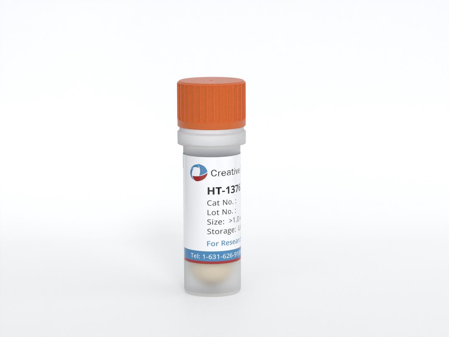
HT-1376
Cat.No.: CSC-C9439L
Species: Homo sapiens (Human)
Source: Bladder
Morphology: epithelial
Culture Properties: monolayer
- Specification
- Background
- Scientific Data
- Q & A
- Customer Review
Tumorigenecity: yes, in mice and hamsters
Isoenzyme: G6PD,B
Production: fibrinolytic activity; interferon
Histopathology: carcinoma; cancer; Grade 3
Note: the line does not contain detectable virus particles, herpesvirus antigens or retrovirus antigens; the cells will grow in soft agar
vWA: 15,18
FGA: 21,22
Amelogenin: X
TH01: 7,9.3
TPOX: 8
CSF1P0: 12
D5S818: 11,12
D13S317: 9,11
D7S820: 9,12
The HT-1376 cell line is a widely utilized human bladder cancer line established from a high-grade (grade 3) primary transitional cell carcinoma (urothelial carcinoma) of a 58-year-old female patient. Because it originates from an untreated primary muscle-invasive tumor, HT-1376 faithfully captures the molecular and biological hallmarks of aggressive bladder cancer without the genomic confounders of prior chemotherapy.
A defining advantage of HT-1376 is its well-characterized p53 pathway deficiency. The cells harbor a homozygous missense mutation in TP53 at codon 245 (G245D), a classic gain-of-function hotspot mutation that abrogates wild-type tumor-suppressor activity and drives genomic instability. This provides a consistent and clinically relevant genetic background for investigating p53-driven tumorigenesis and drug resistance. Phenotypically, HT-1376 cells grow as an adherent monolayer with typical epithelial cobblestone morphology and robustly express urothelial keratins (CK7, CK8, CK18, CK19), confirming lineage fidelity. When implanted subcutaneously into immunodeficient mice, they reliably form moderately differentiated xenograft tumors that recapitulate the infiltrative histology of human muscle-invasive bladder cancer, including stromal invasion.
The combination of a stable epithelial phenotype, precisely defined TP53 mutation, and reproducible in vivo tumorigenicity makes HT-1376 an indispensable platform for dissecting the molecular mechanisms of bladder cancer invasion, evaluating platinum-based and targeted therapeutics, and studying epithelial-mesenchymal transition. Its origin from a primary lesion, rather than a metastatic site, further enhances its relevance as a model for early-stage invasive bladder cancer research.
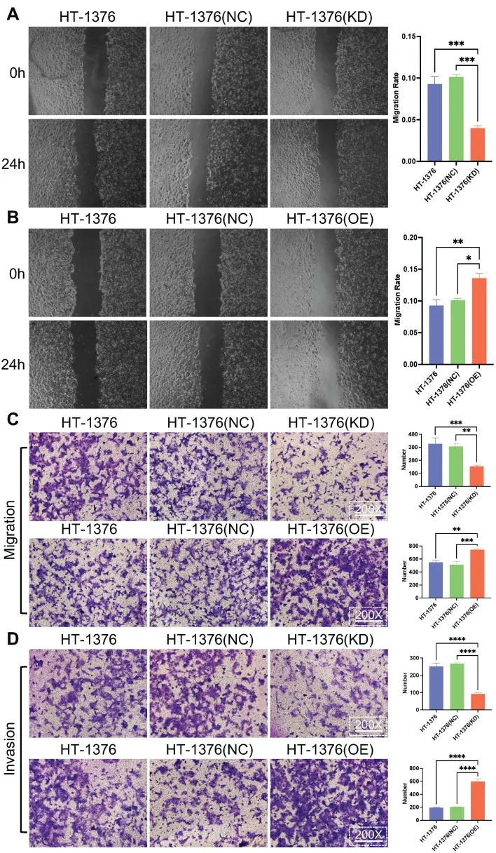
S100A8 Promotes the Proliferation, Migration and Invasion in Bladder Cancer Cells
There is mounting evidence that S100 calcium-binding A8 (S100A8) is involved in inflammation and cancer. However, whether S100A8 promotes the proliferation, invasion and migration of bladder cancer (BC) is still not completely clear. To investigate the influence of S100A8 on the proliferation, migration and invasion of BC.
S100A8 was overexpressed and knocked down in HT-1376 cells via infection with overexpression recombinant lentivirus (OE group) and shRNA‑encoding lentivirus (KD group), respectively. In addition, HT-1376 cells were also infected with a negative control lentivirus as a negative control group (NC group). Cell proliferation was analyzed using the CCK-8 assay and 5-ethynyl-2'-deoxyuridine assay, while migration and invasion capabilities were evaluated via wound healing and Transwell assay.
Compared with the negative control group, the proliferation, migration and invasion of S100A8-overexpressing HT-1376 cells were enhanced, while those of S100A8-knockdown HT-1376 cells were reduced. Consequently, S1008A emerges as a promising diagnostic and therapeutic target for BC.
Ask a Question
Write your own review
- You May Also Need
Description: Contains the unusual type A isoenzyme of glucose-6-phosphate dehydrogenase.
Description: Species: human - male, 44 years old, CaucasianTumorigenecity: yes, in mice and hamstersIsoenzyme: G6PD, BProduction: fibrinolytic activityHistopathology: carcinomaNote: the cells will grow in soft ...
Description: Established from the bladder tumor of an 84-year-old Caucasian woman following transurethral tumor resection in 1977
Description: Species: human - male, 58 years old, Caucasian, SwedishTumorigenecity: noIsoenzyme: Me-2, 1-2; PGM3, 2; PGM1, 1; ES-D, 1; AK-1, 1; G6PD, B; GLO-1Histopathology: carcinoma, transitional cell
Description: Derived from malignant ascitic fluid of a 75-year-old man with urinary bladder carcinoma in 1988; described as being of epithelial origin with morphologically distinct cells (polygonal to ...
Description: Derived from the invasive solid transitional cell carcinoma of the bladder of a 82-year-old Caucasian woman (grade IV, stage C); cell growth described as being inhibited by EGF
- Adipose Tissue-Derived Stem Cells
- Human Neurons
- Mouse Probe
- Whole Chromosome Painting Probes
- Hepatic Cells
- Renal Cells
- In Vitro ADME Kits
- Tissue Microarray
- Tissue Blocks
- Tissue Sections
- FFPE Cell Pellet
- Probe
- Centromere Probes
- Telomere Probes
- Satellite Enumeration Probes
- Subtelomere Specific Probes
- Bacterial Probes
- ISH/FISH Probes
- Exosome Isolation Kit
- Human Adult Stem Cells
- Mouse Stem Cells
- iPSCs
- Mouse Embryonic Stem Cells
- iPSC Differentiation Kits
- Mesenchymal Stem Cells
- Immortalized Human Cells
- Immortalized Murine Cells
- Cell Immortalization Kit
- Adipose Cells
- Cardiac Cells
- Dermal Cells
- Epidermal Cells
- Peripheral Blood Mononuclear Cells
- Umbilical Cord Cells
- Monkey Primary Cells
- Mouse Primary Cells
- Breast Tumor Cells
- Colorectal Tumor Cells
- Esophageal Tumor Cells
- Lung Tumor Cells
- Leukemia/Lymphoma/Myeloma Cells
- Ovarian Tumor Cells
- Pancreatic Tumor Cells
- Mouse Tumor Cells