
IMR-90
Cat.No.: CSC-C9446L
Species: Homo sapiens (Human)
Source: Lung
Morphology: fibroblast
Culture Properties: monolayer
- Specification
- Background
- Scientific Data
- Q & A
- Customer Review
Isoenzyme: G6PD,B
Histopathology: normal
vWA: 16,19
FGA: 25,26
Amelogenin: X
TH01: 8,9.3
TPOX: 8,9
CSF1P0: 11,13
D5S818: 12,13
D13S317: 11,13
D7S820: 9,12
The IMR-90 cell line is a well-characterized human diploid fibroblast strain derived from the normal lung tissue of a 16-week-old female Caucasian fetus, established by W.W. Nichols and colleagues. These adherent cells exhibit typical fibroblast-like morphology and maintain a normal female diploid karyotype (46, XX), providing a physiologically relevant human genetic background that contrasts sharply with the aberrant karyotypes of transformed cancer cell lines.
A defining feature of IMR-90 cells is their finite replicative lifespan. They are capable of achieving approximately 58 population doublings before entering replicative senescence, making them an indispensable in vitro model for studying cellular aging, telomere biology, and senescence-associated pathways. This Hayflick limit enables researchers to investigate the molecular and phenotypic progression from proliferation to senescence in a controlled, human-relevant system.
IMR-90 cells are also highly valued for their viral susceptibility, which is comparable to that of WI-38 and MRC-5 cells. They are susceptible to multiple viruses, including poliovirus, herpes simplex virus, and vaccinia virus, supporting their use in virological studies. Their thoroughly characterized division potential and viral susceptibilities allow them to serve as a standard alternative to WI-38 and other human lung cell strains.
Beyond aging and virology, IMR-90 cells are widely applied in pulmonary fibrosis research, toxicology assays, drug testing, and cytoskeletal dynamics studies. They are amenable to transfection, facilitating genetic and epigenetic investigations. Furthermore, they have been used to generate induced pluripotent stem cells (iPSCs), extending their utility into regenerative medicine.
ICOS+ T Helper 2 Cells Could Promote Pulmonary Fibrosis In Vitro
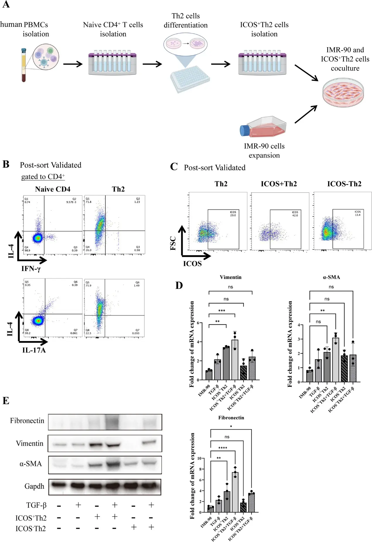
To assess the direct functional effects of ICOS+ Th2 cells on pulmonary fibrosis, Zhu, Huimin, et al. performed in vitro assays using human pulmonary fibroblasts (IMR-90). IMR-90 cells were co-cultured with ICOS+ Th2 cells that had been differentiated in vitro (Fig. 1A) and fibrotic gene expression was measured. Under polarizing conditions and magnetic cell sorting, human naive CD4+ T cells up-regulated a substantial subset coexpressing IL-4 and ICOS (Figs. 1B, 1 C). These cells were co-cultured with IMR-90 at a 1:4 ratio in the presence or absence of suboptimal TGF-β and soluble anti-CD3/anti-CD28 antibodies. After 3 to 4 days in co-culture, ICOS+ Th2 cells upregulated the mRNA expression of Vimentin and Fibronectin in IMR-90 cells. At the same time, in the presence of both TGF-β and ICOS+ Th2 cells, the expression of α-SMA was also significantly increased in IMR-90 cells (Fig. 1D). Similarly, at the protein level, the combined action of TGF-β and Th2 cells can significantly increase the levels of Vimentin, Fibronectin and a-SMA (Fig. 1E). ICOS+ Th2 cells initiate moderate myofibroblast activation, but classical full differentiation and immense matrix deposition strictly require synergistic interaction with TGF-β.
Ask a Question
Write your own review
- You May Also Need
Description: Lung small cell carcinoma producing insulin-like growth factor II. Cell growth is slow.
Description: Lung Cancer-1/squamous. Parent cell line of LC-1/sq-SF, the same patient as LC-F. Cell growth is slow.
Description: Established in 1979 from the bone marrow aspirated from a 55-year-old white man with small cell lung carcinoma prior to treatment; corresponds to NCI-H209; described as expressing neuroendocrine ...
Description: Derived from the lung of a 56-year-old black man with small cell lung carcinoma prior to treatment in 1988
- Adipose Tissue-Derived Stem Cells
- Human Neurons
- Mouse Probe
- Whole Chromosome Painting Probes
- Hepatic Cells
- Renal Cells
- In Vitro ADME Kits
- Tissue Microarray
- Tissue Blocks
- Tissue Sections
- FFPE Cell Pellet
- Probe
- Centromere Probes
- Telomere Probes
- Satellite Enumeration Probes
- Subtelomere Specific Probes
- Bacterial Probes
- ISH/FISH Probes
- Exosome Isolation Kit
- Human Adult Stem Cells
- Mouse Stem Cells
- iPSCs
- Mouse Embryonic Stem Cells
- iPSC Differentiation Kits
- Mesenchymal Stem Cells
- Immortalized Human Cells
- Immortalized Murine Cells
- Cell Immortalization Kit
- Adipose Cells
- Cardiac Cells
- Dermal Cells
- Epidermal Cells
- Peripheral Blood Mononuclear Cells
- Umbilical Cord Cells
- Monkey Primary Cells
- Mouse Primary Cells
- Breast Tumor Cells
- Colorectal Tumor Cells
- Esophageal Tumor Cells
- Lung Tumor Cells
- Leukemia/Lymphoma/Myeloma Cells
- Ovarian Tumor Cells
- Pancreatic Tumor Cells
- Mouse Tumor Cells