
Human Lymphatic Fibroblasts (HLF)
Cat.No.: CSC-7762W
Species: Human
Source: Lymph Node
Cell Type: Fibroblast
- Specification
- Background
- Scientific Data
- Q & A
- Customer Review
Human Lymphatic Fibroblasts (HLF) are a type of primary fibroblast cell line that is derived from human lymphatic tissue. As the name suggests, these cells are specialized to help form and maintain the structure and function of the lymphatic system. Unlike general fibroblasts, HLF are particularly active in producing and remodeling extracellular matrix proteins, such as collagen and fibronectin, which provide support and elasticity to lymphatic vessels.
HLF have a spindle-shaped morphology and are known to be highly responsive to various environmental signals, including inflammatory cytokines and growth factors. This allows them to play a crucial role in lymphangiogenesis, tissue remodeling, and guiding immune cells. These cells can dynamically interact with lymphatic endothelial cells, and can influence lymphatic vessel formation and stability. In some cases, HLF can be more sensitive to fibrotic or inflammatory signals than fibroblasts from other tissues. They are commonly used in research to study lymphatic biology, immune regulation, and disease-associated tissue remodeling. Their unique properties and responsiveness to inflammation make them a valuable model for studying lymphatic fibrosis, tumor-lymphatic interactions, and testing potential therapies aimed at restoring or modulating lymphatic function.
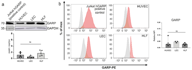
Expression of GARP and Integrins are Produced In Vitro by Human Endothelial Cells and LN Fibroblasts
GARP is expressed on regulatory T lymphocytes (Tregs) in human and murine tumors and activates TGF-β1, causing immunosuppression in tumor-bearing mice. However, its sources and role in lymph nodes (LNs) during metastasis are unclear. LNs, where immune responses occur, contain immune and non-immune cells like blood (BEC)/lymphatic (LEC) endothelial, fibroblastic, and perivascular cells (FRCs).
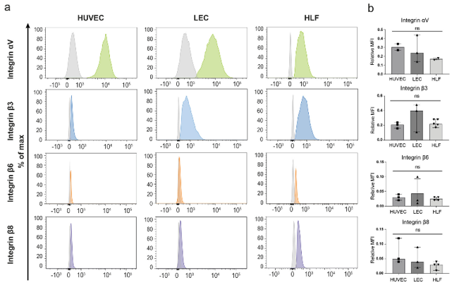
To find which non-immune cells express LRRC32 (the gene for GARP) in human LNs, Rouaud et al. analyzed single-cell RNA sequencing (scRNA-Seq) datasets from human LN samples. This identified LRRC32 expression in endothelial and perivascular cell subpopulations. They then checked GARP protein expression in primary cultures of human umbilical vein endothelial cells (HUVECs), human LECs, and human lymphatic fibroblasts (HLFs). Jurkat cells expressing human GARP served as a positive control. Western blotting showed similar GARP levels in all primary cells (Fig. 1a and Fig. 2). Flow cytometry confirmed GARP on the cell surface of HUVECs, LECs, and HLFs (Figu. 1b). Since integrins αVβ6 and αVβ8 can activate latent TGF-β1 presented by GARP, we checked their presence on these cells, along with the common integrin αVβ3. Flow cytometry detected significant levels of αV and β3 subunits in all three primary cell cultures (Fig. 2). β6 and β8 subunits were also present. Thus, in basal culture conditions, GARP, αVβ6, and αVβ8 are co-expressed on the surface of primary HUVEC, LEC, and HLF cultures.
Ask a Question
Write your own review
Description: NPB CD4+ Helper T cells are negatively isolated from mononuclear cells using an indirect immunomagnetic CD4+ T cell labeling system.
Description: Cord Blood-Pan T cells are negatively isolated from mononuclear cells, using an indirect immunomagnetic Pan T labeling system.

Description: First, NPB-CD4+ T Cells are negatively isolated using immunomagnetic CD4+ isolation kit from mononuclear cells. Next, CD45RO MicroBeads are used to deplete the CD45RO+ cell population, leaving an ...
Description: Chronic Lymphoid Leukemia-PB-CD19+/CD5+ B cells are isolated from Chronic Lymphoid Leukemia bone marrow mononuclear cells using a direct immunomagnetic CD19+ Micro- Bead labeling system. Available in ...

Description: Untouched NPB-CD19+ B Cells are negatively isolated from mononuclear cells using an indirect immunomagnetic labeling system.

Description: NPB-CD19+/CD27+ memory B cells are negatively isolated from Mononuclear Cells using immunomagnetic isolation system to deplete non-memory B cells