
SW-1710
Cat.No.: CSC-C0463
Species: Homo sapiens (Human)
Morphology: epithelial-like, elongated cells growing adherently as monolayers
Culture Properties: monolayer
- Specification
- Background
- Scientific Data
- Q & A
- Customer Review
Immunology: cytokeratin +, cytokeratin-7 -, cytokeratin-8 +, cytokeratin-17 -, cytokeratin-18 +, desmin -, endothel -, EpCAM -, GFAP -, neurofilament -, vimentin (+)
Viru
SW-1710 cells are human cancer cells, used in cancer research. They were originally developed from a primary urothelial (transitional cell) carcinoma of the urinary bladder and are described as a continuous cell line which was isolated from the tumor of an 84-year-old Caucasian female in 1977 after transurethral resection. The cells are epithelial-like, elongated, and adhere to plastic to form monolayers when maintained in vitro. They do not have DNA microsatellite instability, and have a near normal doubling time of approximately 25-32 hours when maintained in culture.
SW-1710 cells have a homozygous mutation in TP53, specifically p.Arg273Cys. The cells are found in several large collections of cancer cell lines used for research including the Cancer Dependency Map, the Cancer Cell Line Encyclopedia (CCLE) and the COSMIC cell line project. SW-1710 has been used as a model of human bladder cancer in many studies to examine tumor biology, determine function of genes of interest, test sensitivity to drugs and chemotherapy, and microenvironmental responses such as hypoxia induction. SW-1710 cells have been used to determine how bladder carcinoma cells respond to hypoxia.
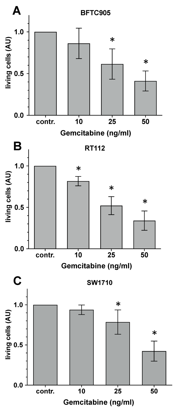
Evaluation of the Cytotoxic Capacity of Gemcitabine
Bladder cancer is one of the most frequent tumors and gemcitabine is a chemotherapeutic agent used for off-label intravesical instillation therapy. Sturm et al. investigated whether the addition of blue light (453 nm) and riboflavin to gemcitabine treatment could increase cytotoxicity induced by gemcitabine alone in bladder cancer cell lines (BFTC-905, SW-1710, RT-112).
Cells were incubated with either 10, 25 or 50 ng/mL gemcitabine for 24 hours. Cell viability was determined by CTB assay. As shown in Figure 1, increasing concentrations of gemcitabine induced concentration-dependent cytotoxicity. Treatment with 10 ng/mL gemcitabine induced significant toxicity in RT-112 cells only (-10% viability) (Fig. 1B). There were no significant differences in viability in BFTC-905 cells (Fig. 1A) or SW-1710 cells (Fig. 1C) treated with 10 ng/mL gemcitabine. Treatment with 25 ng/mL gemcitabine induced significant toxicity in all three cell lines. RT-112 cells were most affected by gemcitabine treatment (40-60% loss), followed by BFTC-905 cells (30-60%) and SW-1710 cells (10-30%). The highest amount of toxicity was achieved in all cell lines treated with 50 ng/mL gemcitabine. Toxicity values ranged from 50-80% and there were no significant differences between any of the cell lines.
Ask a Question
Write your own review
- You May Also Need

Description: Contains the unusual type A isoenzyme of glucose-6-phosphate dehydrogenase.
Description: Species: human - male, 44 years old, CaucasianTumorigenecity: yes, in mice and hamstersIsoenzyme: G6PD, BProduction: fibrinolytic activityHistopathology: carcinomaNote: the cells will grow in soft ...
Description: Species: human - female, 58 years old, CaucasianTumorigenecity: yes, in mice and hamstersIsoenzyme: G6PD,BProduction: fibrinolytic activity; interferonHistopathology: carcinoma; cancer; Grade 3Note: ...
Description: Species: human - male, 58 years old, Caucasian, SwedishTumorigenecity: noIsoenzyme: Me-2, 1-2; PGM3, 2; PGM1, 1; ES-D, 1; AK-1, 1; G6PD, B; GLO-1Histopathology: carcinoma, transitional cell

Description: Derived from malignant ascitic fluid of a 75-year-old man with urinary bladder carcinoma in 1988; described as being of epithelial origin with morphologically distinct cells (polygonal to ...
Description: Derived from the invasive solid transitional cell carcinoma of the bladder of a 82-year-old Caucasian woman (grade IV, stage C); cell growth described as being inhibited by EGF
- Adipose Tissue-Derived Stem Cells
- Human Neurons
- Mouse Probe
- Whole Chromosome Painting Probes
- Hepatic Cells
- Renal Cells
- In Vitro ADME Kits
- Tissue Microarray
- Tissue Blocks
- Tissue Sections
- FFPE Cell Pellet
- Probe
- Centromere Probes
- Telomere Probes
- Satellite Enumeration Probes
- Subtelomere Specific Probes
- Bacterial Probes
- ISH/FISH Probes
- Exosome Isolation Kit
- Human Adult Stem Cells
- Mouse Stem Cells
- iPSCs
- Mouse Embryonic Stem Cells
- iPSC Differentiation Kits
- Mesenchymal Stem Cells
- Immortalized Human Cells
- Immortalized Murine Cells
- Cell Immortalization Kit
- Adipose Cells
- Cardiac Cells
- Dermal Cells
- Epidermal Cells
- Peripheral Blood Mononuclear Cells
- Umbilical Cord Cells
- Monkey Primary Cells
- Mouse Primary Cells
- Breast Tumor Cells
- Colorectal Tumor Cells
- Esophageal Tumor Cells
- Lung Tumor Cells
- Leukemia/Lymphoma/Myeloma Cells
- Ovarian Tumor Cells
- Pancreatic Tumor Cells
- Mouse Tumor Cells