
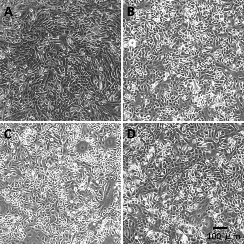
C57BL/6 Mouse Primary Corneal Epithelial Cells
Cat.No.: CSC-C4272X
Species: Mouse
Source: Cornea; Eye
Cell Type: Epithelial Cell
- Specification
- Background
- Scientific Data
- Q & A
- Customer Review
Mouse Primary Corneal Epithelial Cells can be used in assays of cell to cell adhesion and migration. Standard biochemical procedures performed with epithelial cell cultures include RT-PCR, Western blotting, immunoprecipitation, immunofluorescent staining or immunofluorescent flow cytometry or generating cell derivatives for desired research applications.
C57BL/6 Mouse Primary Corneal Epithelial Cells are primary cells isolated from the corneal epithelium of C57BL/6 mice, which are one of the most commonly used inbred strains for biomedical research. The corneal epithelium is a stratified, non-keratinized epithelial tissue that covers the outer surface of the cornea. It plays a crucial role in maintaining corneal transparency, barrier function, and homeostasis of the ocular surface. Primary corneal epithelial cells from this strain are characterized by a native-like morphology, gene expression, and functional properties similar to those found in vivo, making them a relevant model system for mechanistic and translational studies.
The cells express epithelial markers such as cytokeratins (e.g., K12, K14), E-cadherin, and tight junction proteins like ZO-1 and occludin, reflecting their barrier-forming properties. These cells are used to study corneal development, epithelial differentiation, wound healing, and regeneration. They are also employed as in vitro models for investigating ocular surface inflammation, dry eye disease, microbial infection, and corneal toxicity caused by drugs or environmental stressors.
Effects of 1,25-Vitamin D3 and 24,25-Vitamin D3 on Corneal Nerve Regeneration in Diabetic Mice
This study examined how vitamin D affects corneal nerve regeneration after epithelial injury in diabetic mice. Diabetes, VDR knockout (VDR KO), and vitamin D deficiency (VDD) significantly slowed nerve regeneration. However, applying 1,25 Vit D and 24,25 Vit D topically sped up this process.
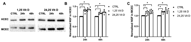
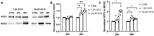
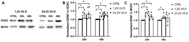
In human corneal epithelial cells (HCECs) and mouse corneal epithelial cells (MCECs), NGF protein expression increased after 1,25 Vit D and 24,25 Vit D exposure (Fig. 1). NTF3 protein expression in HCECs significantly rose 48 hours after exposure to 1,25 Vit D and 24,25 Vit D, but not at 24 hours. In MCECs, NTF3 protein expression significantly increased 24 hours after 24,25 Vit D exposure and 48 hours after both 1,25 Vit D and 24,25 Vit D exposure (Fig. 2). GDNF protein expression in HCECs significantly increased 24 and 48 hours after 1,25 Vit D and 24,25 Vit D exposure. In MCECs, GDNF protein expression significantly increased 24 hours after 1,25 Vit D and 24,25 Vit D exposure, and 48 hours after 1,25 Vit D exposure (Fig. 3).
Ask a Question
Write your own review
Description: C57BL/6-GFP Mouse Skeletal Muscle Microvascular Endothelial Cells from Creative Bioarray are isolated from C57BL/6-Tg (CAG-EGFP) 1Osb/J mouse skeletal muscle tissue of pathogen-free laboratory mice. ...

Description: eNOS KO Mouse Stomach Epithelial Cells from Creative Bioarray are isolated from stomach tissue of pathogen-free laboratory mice. eNOS KO Mouse Stomach Epithelial Cells are grown in a T25 tissue ...

Description: eNOS KO Mouse Liver Fibroblasts from Creative Bioarray are isolated from liver tissue of pathogen-free laboratory mice. eNOS KO Mouse Liver Fibroblasts are grown in T75 tissue culture flasks ...

Description: C57BL/6-GFP Mouse Corneal Epithelial Cells from Creative Bioarray are isolated from C57BL/6-GFP-Tg(CAG-EGFP)1Osb/J mouse corneal tissue of pathogen-free laboratory mice. C57BL/6-GFP Mouse Corneal ...

Description: BALB/c Mouse Retinal Microvascular Endothelial Cells from Creative Bioarray are isolated from retinal tissue of pathogen-free laboratory mice. BALB/c Mouse Retinal Microvascular Endothelial Cells are ...