
Immortalized Rat Odontoblast Cells
Cat.No.: CSC-I9246L
Species: Rattus norvegicus
Source: Molar tooth germs
Culture Properties: Adherent
- Specification
- Background
- Scientific Data
- Q & A
- Customer Review
Note: Never can cells be kept at -20 °C.
2) in vitro mineralization activity confirmed by Von Kossa staining;
3) in vivo mineralization activity verified bysubcutaneous and intramuscular implantation.
Immortalized Rat Odontoblast Cells are primary cell cultures which are mesenchymal cells isolated from either rat dental pulp or dental papilla that have been genetically altered or selected for continued proliferation in vitro.
Typically, immortalized rat odontoblast cells are adherent with spindle-shaped to columnar appearance. They may also be polarized in a manner similar to that seen in primary odontoblasts that line the pulp chamber. Immortalized rat odontoblast cells express odontoblast-specific markers such as DSPP, dentin matrix protein 1 (DMP1), alkaline phosphatase (ALP), and type I collagen, which they utilize during production and mineralization of dentin matrix. Similar to primary odontoblasts, immortalized rat odontoblast cells will also mineralize to form mineralized nodules and are responsive to bone morphogenetic proteins (BMPs), transforming growth factor-β (TGF-β), and Wnt signaling. Immortalized rat odontoblast cells are commonly used in research surrounding tooth development, dentinogenesis, and tooth tissue regeneration due to their stable phenotype and continued ability to differentiate along the odontoblast lineage.
Low-dose AgNPs Exhibit Minimal Cytotoxicity and Restore Cell Viability in LPS-challenged Immortalized Rat Odontoblast Cells
Silver nanoparticles (AgNPs) are commonly used nanomaterials in biomedicine due to their antimicrobial activity. However, the effect of AgNPs on odontogenic cells subjected to inflammation remains to be elucidated. Wang et al. explored the effect of low dose AgNPs on mineralization and inflammatory response on lipopolysaccharide (LPS)-stimulated immortalized rat odontoblast cells (MDPC-23) through the PI3K/Akt-NF-κB pathway.
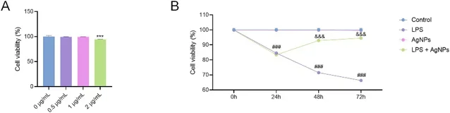
First, they determined a non-toxic concentration by treating MDPC-23 cells with AgNPs at doses of 0-2 μg/mL for 24 h. Cell viability assays demonstrated that cells treated with 0.5 and 1 μg/mL AgNPs had similar viability to controls while cell viability significantly decreased when treated with 2 μg/mL AgNPs (Fig. 1A). As a result, 1 μg/mL AgNPs were used for subsequent experiments. Cells were then treated under inflammatory conditions through four groups (control, LPS, AgNPs, LPS + AgNPs) for up to 72 h. They found that LPS significantly decreased cell viability beginning at 24 h reaching approximately 70% viability at 72 h compared to controls (Fig. 1B). AgNPs alone had no effect on cell viability, however co-treatment with LPS and AgNPs significantly improved cell survival at 48 h and 72 h. These results demonstrated that low dose AgNPs are not cytotoxic and can attenuate proliferative deficits caused by LPS in MDPC-23 cells.
Ask a Question
Write your own review

Description: Immortalized Mouse Primary Artery Fibroblasts-GFP provided by Creative Bioarray have been developed by immortalizing mouse artery fibroblasts with SV40 Large T antigen and transfecting with tGFP. The ...
Description: Immortalized MHC II -/- Mouse Macrophage Cells (C2D) is derived from the knockout mice negative for MHCII molecule. They are stable in culture and survived through crisis in which they spontaneously ...
Description: The bone morphogenetic proteins (BMPs) produced by calvarial cells are crucial in osteoblastic differentiation and bone regeneration, however the limited lifespan of primary cells diminishes their ...
Description: The Immortalized Mouse Atrioventricular Cushion Mesenchymal Cells (tsA58-AVM) were derived from atrioventricular (AV) cushions of H-2Kb -tsA58 embryos at E9.5 and were conditionally immortalized in ...
Description: IDG-SW3 represent a non-homogenous population progressing from early osteoblasts to late osteocytic. These cells express functional SV40 large T antigen that is induced in the presence of IFN γ under ...
Description: The Immortalized Mouse Spleen Dendritic Cell (SRDC) line is functionally and phenotypically similar to dendritic cells; specifically, in its antigen presentation, T cell priming, and dendritic cell ...
- Adipose Tissue-Derived Stem Cells
- Human Neurons
- Mouse Probe
- Whole Chromosome Painting Probes
- Hepatic Cells
- Renal Cells
- In Vitro ADME Kits
- Tissue Microarray
- Tissue Blocks
- Tissue Sections
- FFPE Cell Pellet
- Probe
- Centromere Probes
- Telomere Probes
- Satellite Enumeration Probes
- Subtelomere Specific Probes
- Bacterial Probes
- ISH/FISH Probes
- Exosome Isolation Kit
- Human Adult Stem Cells
- Mouse Stem Cells
- iPSCs
- Mouse Embryonic Stem Cells
- iPSC Differentiation Kits
- Mesenchymal Stem Cells
- Immortalized Human Cells
- Immortalized Murine Cells
- Cell Immortalization Kit
- Adipose Cells
- Cardiac Cells
- Dermal Cells
- Epidermal Cells
- Peripheral Blood Mononuclear Cells
- Umbilical Cord Cells
- Monkey Primary Cells
- Mouse Primary Cells
- Breast Tumor Cells
- Colorectal Tumor Cells
- Esophageal Tumor Cells
- Lung Tumor Cells
- Leukemia/Lymphoma/Myeloma Cells
- Ovarian Tumor Cells
- Pancreatic Tumor Cells
- Mouse Tumor Cells