
Immortalized Mouse Articular Chondrocytes (iMAC)
Cat.No.: CSC-I9225L
Species: Mus musculus
Source: Tibial Plateau, Femoral Head, and Femoral Condyles
Morphology: Polygonal
Culture Properties: Adherent
- Specification
- Background
- Scientific Data
- Q & A
- Customer Review
Note: Never can cells be kept at -20 °C.
2) Col 1al immunohistochemical staining preformed;
3) Western blot used to detect SV40 large T antigen presence;
4) Alcian blue staining analysis for chondroid matrix production.
Immortalized Mouse Articular Chondrocytes, commonly abbreviated iMAC, are immortalized cells derived from mouse articular cartilage chondrocytes. They were originally isolated and immortalized in order to develop a cell line that had continuous proliferation potential without losing important phenotypic or functional characteristics of normal chondrocytes. Typically, chondrocytes harvested from articular/cartilage joints of mice are used to generate iMAC cells.
Immortalized mouse articular chondrocytes in vitro have an adherent growth pattern with polygonal to rounded morphology typical of articular chondrocytes with some fibroblastic morphology that can occur with long-term passaging. Importantly, they continue to express type II collagen (COL2A1), aggrecan (ACAN), and SOX9. Under certain culture conditions, such as 3D culture or chondrogenic media, iMAC cells have been shown to deposit cartilage-like matrix and form proteoglycan-rich deposits. Because of these phenotypic characteristics, iMAC cells are often used to study cartilage homeostasis, ECM metabolism, and chondrocyte differentiation and dedifferentiation. These cells are commonly used to study the cellular mechanisms that contribute to cartilage degeneration and osteoarthritis (OA) such as inflammation (NF-κB, MAPK, Wnt/β-catenin signaling) and catabolic processes involving matrix degrading enzymes (MMPs and ADAMTS). These cells are also commonly used to study mechanical stress, oxidative stress, and cytokine-induced damage.
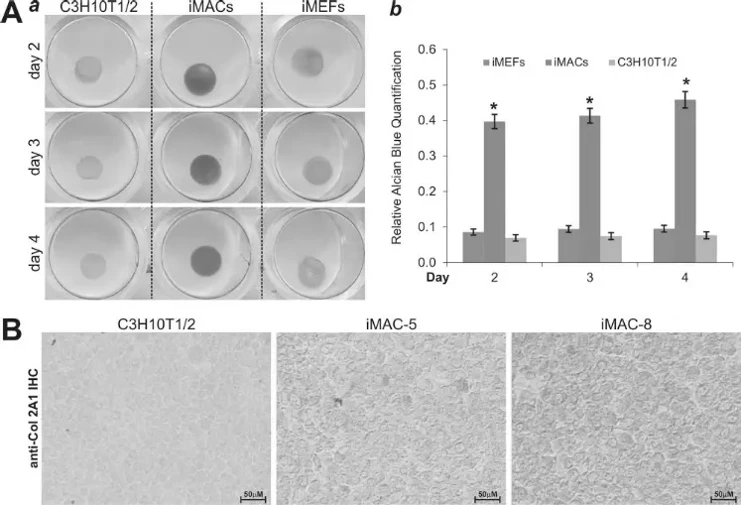
iMACs can Effectively Produce Chondroid Matrix in Micromass Culture
Cartilage tissue engineering holds great promise for treating cartilaginous Cartilage tissue engineering offers significant potential for treating degenerative and traumatic cartilaginous injuries, but obtaining sufficient chondrocytes remains challenging due to the limited proliferative capacity of primary cells. Lamplot et al. investigated whether reversibly immortalized mouse articular chondrocytes (iMACs) maintain long-term proliferative capability while preserving chondrogenic phenotype.
They assessed chondroid matrix production using micromass culture. Highly concentrated iMACs and control MSCs (C3H/10T1/2 and iMEFs) were seeded to form micromasses. Alcian blue staining demonstrated significantly increased chondroid matrix production in iMAC clones compared to MSC controls (Fig. 1A). Immunohistochemical analysis further revealed higher Col2a1 expression in iMAC micromasses versus C3H/10T1/2 cells (Fig. 1B). These results confirm that iMACs retain chondrogenic phenotype in vitro.
Ask a Question
Write your own review
Description: Immortalized Mouse Primary Artery Fibroblasts-GFP provided by Creative Bioarray have been developed by immortalizing mouse artery fibroblasts with SV40 Large T antigen and transfecting with tGFP. The ...

Description: Immortalized MHC II -/- Mouse Macrophage Cells (C2D) is derived from the knockout mice negative for MHCII molecule. They are stable in culture and survived through crisis in which they spontaneously ...
Description: The bone morphogenetic proteins (BMPs) produced by calvarial cells are crucial in osteoblastic differentiation and bone regeneration, however the limited lifespan of primary cells diminishes their ...
Description: The Immortalized Mouse Atrioventricular Cushion Mesenchymal Cells (tsA58-AVM) were derived from atrioventricular (AV) cushions of H-2Kb -tsA58 embryos at E9.5 and were conditionally immortalized in ...
Description: IDG-SW3 represent a non-homogenous population progressing from early osteoblasts to late osteocytic. These cells express functional SV40 large T antigen that is induced in the presence of IFN γ under ...
Description: The Immortalized Mouse Spleen Dendritic Cell (SRDC) line is functionally and phenotypically similar to dendritic cells; specifically, in its antigen presentation, T cell priming, and dendritic cell ...
- Adipose Tissue-Derived Stem Cells
- Human Neurons
- Mouse Probe
- Whole Chromosome Painting Probes
- Hepatic Cells
- Renal Cells
- In Vitro ADME Kits
- Tissue Microarray
- Tissue Blocks
- Tissue Sections
- FFPE Cell Pellet
- Probe
- Centromere Probes
- Telomere Probes
- Satellite Enumeration Probes
- Subtelomere Specific Probes
- Bacterial Probes
- ISH/FISH Probes
- Exosome Isolation Kit
- Human Adult Stem Cells
- Mouse Stem Cells
- iPSCs
- Mouse Embryonic Stem Cells
- iPSC Differentiation Kits
- Mesenchymal Stem Cells
- Immortalized Human Cells
- Immortalized Murine Cells
- Cell Immortalization Kit
- Adipose Cells
- Cardiac Cells
- Dermal Cells
- Epidermal Cells
- Peripheral Blood Mononuclear Cells
- Umbilical Cord Cells
- Monkey Primary Cells
- Mouse Primary Cells
- Breast Tumor Cells
- Colorectal Tumor Cells
- Esophageal Tumor Cells
- Lung Tumor Cells
- Leukemia/Lymphoma/Myeloma Cells
- Ovarian Tumor Cells
- Pancreatic Tumor Cells
- Mouse Tumor Cells