
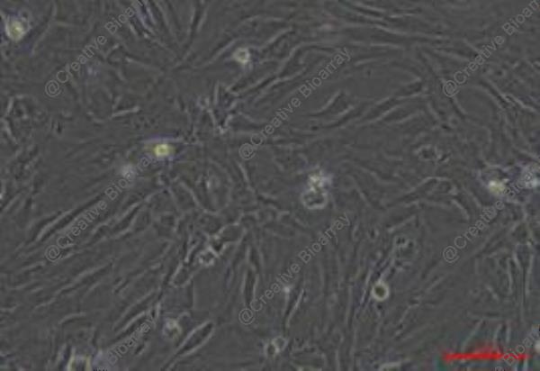
Immortalized Human Skin Fibroblast-SV40
Cat.No.: CSC-I9011L
Species: homo sapiens
Source: Skin
Morphology: Bipolar
Culture Properties: Adherent
- Specification
- Background
- Scientific Data
- Q & A
- Customer Review
- Documents
Note: Never can cells be kept at -20 °C.
CIK-HT003 HT® Lenti-SV40T Immortalization Kit
Skin fibroblasts are crucial to skin biology, playing a key role in extracellular matrix production, wound healing, and skin regeneration. Traditionally, primary skin fibroblasts have been considered the gold-standard model system for studying skin biology, however, donor variability and the limited lifespan of primary cells restrict their potential.
Immortalized Human Skin Fibroblast-SV40 overcomes the limitations of primary cells while retaining the functional characteristics of primary cells. This novel cell line provides a robust and reproducible in vitro cell model for skin disease, skin aging, wound healing, cosmetic, and toxicology studies.
SV40-immortalized human skin fibroblasts exhibit distinct fibroblast-like morphology, express TE-7, and are capable of differentiating into myofibroblasts. Similar to the parental primary cells, SV40-immortalized human skin fibroblasts respond to drug treatment, such as Chlorhexidine (CHX), in a time- and dose-dependent manner. Furthermore, when SV40-immortalized human skin fibroblasts are co-cultured with primary or immortalized keratinocytes (CSC-C4005X or CSC-I9096L), 3D skin structures were formed in vitro.
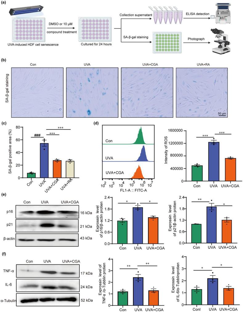
Screening of Active Molecules to Prevent UVA-Induced Senescence of HDF Cells
Cellular senescence plays a critical role in repeated ultraviolet (UV) exposure-induced skin photoaging. Currently, from the perspective of regulating senescent cells, potent compounds or reliable protein targets that could effectively prevent skin photoaging have not yet been reported.
Candidate compounds against UVA-induced photoaging were screened from 27 natural products at a concentration of 10 μM. The immortalized human dermis skin fibroblasts (HDF) were treated with UVA irradiation for 30 min to induce senescence, and 10 μM natural products were added to the medium and incubated with the cells for 24 h, as Fig. 1a depicted. Senescent cells were characterized by elevated SA-β-gal activity, SASP secretion and increased p16 and p21 protein expression.
The results indicated that chlorogenic acid (CGA) could effectively reduce the proportion of SA-β-gal positive cells induced by UVA treatment. The ELISA data confirmed that CGA exhibited the best biologic activity in inhibiting the senescence of HDF, supported by the decreased TNF-α and IL-6 secretion in treated groups. Furthermore, CGA is just as effective as rapamycin (RA, a positive drug that suppress senescence, concentration in 5 nM) in preventing UVA-induced senescence of HDF cells (Fig. 1b, c). Flow cytometry results showed that CGA also inhibited UVA-induced elevation of ROS level (Fig. 1d). And western blot analysis further showed that CGA could effectively inhibit the expression levels of p16 and p21 proteins in UVA-induced HDF cells (Fig. 1e). Moreover, CGA can also significantly suppressed the expression levels of inflammatory factors TNF-α and IL-6 proteins in UVA-treated HDF cells (Fig. 1f). In conclusion, these results demonstrated that CGA effectively prevented UVA induced senescence of HDF in vitro.
Thaw frozen tubes containing 1 mL of cell suspension by rapid shaking in a 37℃ water bath, add 5 mL of culture medium and mix well. Centrifuge at 1000 rpm for 5 minutes, discard the supernatant, replenish 4-6 mL of complete medium and blow well. All cell suspensions are then added to culture flasks and incubated overnight (or cell suspensions are added to 6 cm dishes) and incubated overnight. The next day, the solution is changed and the cell density is checked.
Immortalized Human Skin Fibroblasts-SV40 are ideal for studying skin biology, wound healing, fibrosis, drug screening, and cosmetic testing. They provide a reliable and scalable model for testing and research in dermatological sciences.
Yes, we offer custom cell line development tailored to meet unique research requirements. Please contact us to discuss your specific needs and learn more about our custom solutions.
Ask a Question
Average Rating: 5.0 | 1 Scientist has reviewed this product
Professional
Creative Bioarray is a professional in the field of cells and has helped me a lot.
12 Feb 2021
Ease of use
After sales services
Value for money
Write your own review
Description: Nasal epithelial cells form the outermost protective layer against environmental factors. They clean, humidify, and warm inhaled air. They produces mucus, which binds particles that are subsequently ...

Description: Immortalized Human Corneal Epithelial Cells-SV40 have been obtained immortalizing Human Corneal Epithelial Cells with Lenti-SV40 Lentivirus. Immortalized cells were controlled passaging side by side ...
Description: Immortalized Human Lymphatic Endothelial Cells-SV40 were developed from human tissues transduced with a lentiviral expression vector containing the SV40T gene. The cell line was continuously cultured ...
Description: Immortalized Human Retinal Pigment Epithelial Cells were isolated from neonatal human globes and spontaneously immortalized in culture bypassing crisis. They retained typical morphology of the ...
- Adipose Tissue-Derived Stem Cells
- Human Neurons
- Mouse Probe
- Whole Chromosome Painting Probes
- Hepatic Cells
- Renal Cells
- In Vitro ADME Kits
- Tissue Microarray
- Tissue Blocks
- Tissue Sections
- FFPE Cell Pellet
- Probe
- Centromere Probes
- Telomere Probes
- Satellite Enumeration Probes
- Subtelomere Specific Probes
- Bacterial Probes
- ISH/FISH Probes
- Exosome Isolation Kit
- Human Adult Stem Cells
- Mouse Stem Cells
- iPSCs
- Mouse Embryonic Stem Cells
- iPSC Differentiation Kits
- Mesenchymal Stem Cells
- Immortalized Human Cells
- Immortalized Murine Cells
- Cell Immortalization Kit
- Adipose Cells
- Cardiac Cells
- Dermal Cells
- Epidermal Cells
- Peripheral Blood Mononuclear Cells
- Umbilical Cord Cells
- Monkey Primary Cells
- Mouse Primary Cells
- Breast Tumor Cells
- Colorectal Tumor Cells
- Esophageal Tumor Cells
- Lung Tumor Cells
- Leukemia/Lymphoma/Myeloma Cells
- Ovarian Tumor Cells
- Pancreatic Tumor Cells
- Mouse Tumor Cells