

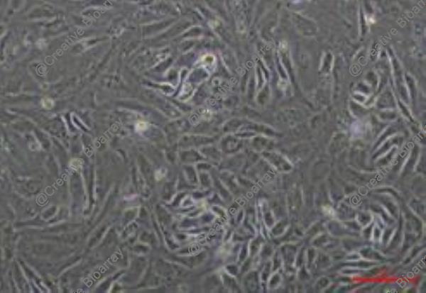
Immortalized Human Hepatic Sinusoidal Endothelial Cells
Cat.No.: CSC-I9188L
Species: Homo sapiens
Source: Liver
Morphology: Cobblestone-like
Culture Properties: Adherent
- Specification
- Background
- Scientific Data
- Q & A
- Customer Review
- Documents
SEC are microvascular endothelial cells with a unique phenotype reminiscent of dendritic cells and a unique function as antigenpresenting cells for CD4+ T cells. Thus, SEC represent a new type of organ resident "nonprofessional" antigenpresenting cell that appears to be involved in the local control of the immune response and the induction of immune tolerance in the liver. Primary SEC express well characterized surface receptors and differ morphologically and metabolically from largevessel endothelia. It has reported that SEC are dynamic regulators of porosity that respond rapidly and locally to environmental zonal stimuli during liver regeneration.
Note: Never can cells be kept at -20°C.
The liver sinusoidal endothelium functions as a highly specialized vascular interface, where liver sinusoidal endothelial cells (LSECs) form a unique fenestrated barrier that lines the hepatic sinusoids and comprises the majority of endothelial cells within the liver. LSECs are distinguished by their lack of an organized basement membrane and the presence of transcellular fenestrae, which collectively ensure high vascular permeability and facilitate efficient bidirectional exchange of nutrients and solutes between the sinusoidal blood and the space of Disse. Beyond their role as a passive filtration barrier, LSECs actively participate in blood clearance via receptor-mediated endocytosis, immune surveillance, and the regulation of vascular tone. However, primary LSECs are notoriously difficult to obtain due to the scarcity of suitable liver resections, and their use for in vitro investigations is compromised by poor cell yields and a limited proliferation capacity, which severely restricts long-term mechanistic studies.
To overcome these limitations, human LSECs have been immortalized through stable introduction of immortalizing genes, including Simian Virus 40 large T antigen (SV40T), human telomerase reverse transcriptase (hTERT), or both. The immortalized cells retain key endothelial characteristics, expressing classic markers such as von Willebrand Factor (vWF), CD31, VE-cadherin, and stabilin family markers. They maintain functional LSEC features, including endocytosis of acetylated low-density lipoprotein (Ac-LDL), tube formation capacity, and immune reactivity. More importantly, they preserve the characteristic fenestrated morphology and Weibel-Palade bodies, as confirmed by scanning and transmission electron microscopy, while offering sustained expansion and experimental reproducibility essential for high-throughput studies.
Collectively, immortalized human LSECs provide an unlimited, consistent, and phenotypically faithful in vitro model for investigating LSEC physiology and pathobiology-including liver regeneration, infection, ischemia-reperfusion injury, and drug-induced hepatotoxicity-circumventing the inherent limitations of primary cell cultures.
Inhibition of PCSK9 Attenuates Liver Endothelial Cell Activation Induced by Colorectal Cancer Stem Cells During Liver Metastasis
Colorectal cancer (CRC) is among the most prevalent and lethal cancers globally, with liver metastasis representing the leading cause of CRC-related mortality. Proprotein convertase subtilisin/kexin type 9 (PCSK9) has recently gained attention due to its overexpression in colorectal tumor tissues and its potential role in driving metastatic progression. This aims to investigate the involvement of PCSK9 in the liver metastatic niche, focusing on its effects on liver sinusoidal endothelial cells (LSECs), key components of the liver microenvironment.
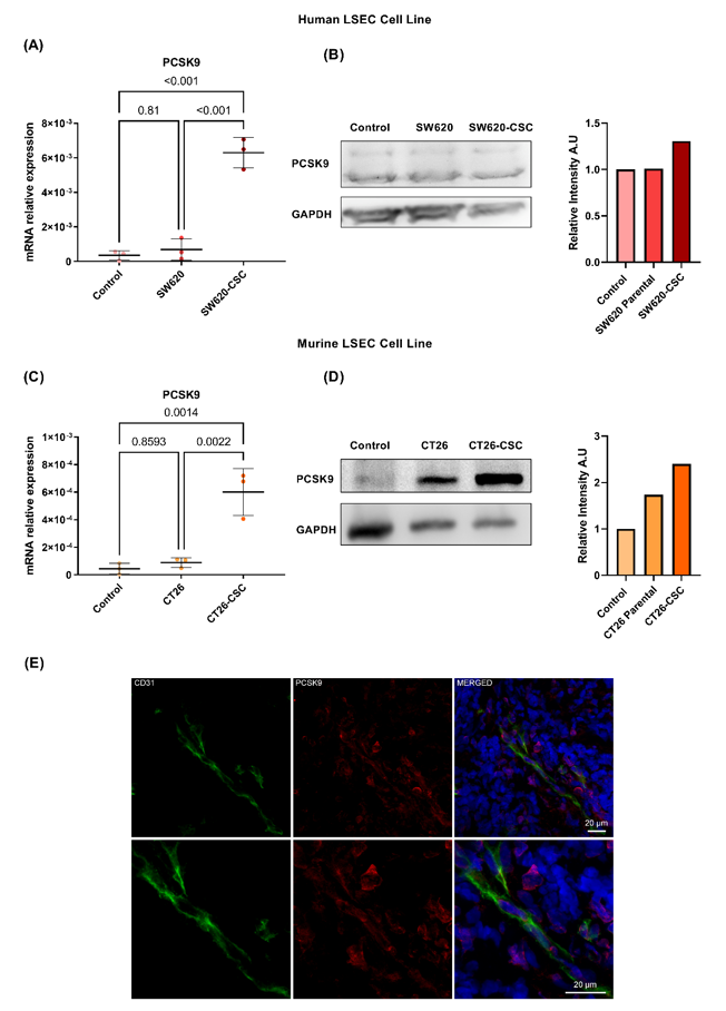
LSECs were stimulated with conditioned media derived from differentiated colorectal cancer cells and cancer stem cells (CSCs), the latter generated by reprogramming SW620 and CT26 cell lines. RNA sequencing was used to profile gene expression in LSECs. PCSK9 mRNA and protein levels were quantified by qPCR and Western blotting, respectively. PCSK9 expression in CRC liver metastases was evaluated by immunofluorescent staining.
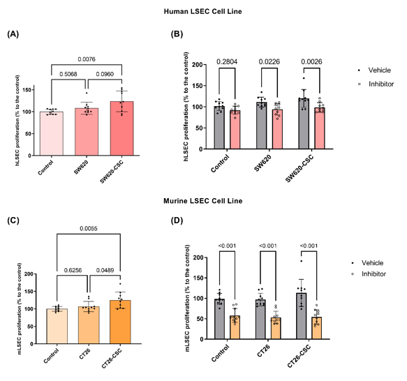
PCSK9 was detected in both human and murine LSECs and significantly upregulated following exposure to CSC-conditioned media. Immunofluorescent staining confirmed PCSK9 expression in LSECs within CRC liver metastases. Total RNA sequencing revealed that a pre-treatment of LSECs with the PCSK9 inhibitor PF-06446864 prior to CSC stimulation seems to reduce the expression of microRNAs linked to cell migration and proliferation. Functional assays demonstrated that CSC-conditioned media enhanced LSEC proliferation and migration, effects reversed by PCSK9 inhibition. These findings highlight PCSK9 as a potential therapeutic target in colorectal liver metastasis.
These are human hepatic sinusoidal endothelial cells that have been genetically modified to go beyond 30 passages, providing a stable and consistent in vitro model for research on liver endothelial functions and diseases.
Immortalized Human Hepatic Sinusoidal Endothelial Cells (Cat No.: CSC-I9188L) are perfect for studying liver microcirculation, endothelial cell function within the liver, liver fibrosis, drug metabolism, toxicology studies, and vascular biology in hepatic contexts.
Unlike primary hepatic endothelial cells, which have a limited lifespan and batch variability, these immortalized cells offer a reproducible and stable model, facilitating long-term studies and consistent experimental outcomes.
Ask a Question
Write your own review
 Immortalized Human Hepatic Sinusoidal Endothelial Cells
Description: Nasal epithelial cells form the outermost protective layer against environmental factors. They clean, humidify, and warm inhaled air. They produces mucus, which binds particles that are subsequently ...

Description: Immortalized Human Splenic Endothelial Cells-SV40 have been obtained immortalizing Human Splenic Endothelial Cells with SV40LT expressing lentiviral particles. Immortalized cells were controlled ...
Description: Immortalized Human Corneal Epithelial Cells-SV40 have been obtained immortalizing Human Corneal Epithelial Cells with Lenti-SV40 Lentivirus. Immortalized cells were controlled passaging side by side ...
Description: Immortalized Human Lymphatic Endothelial Cells-SV40 were developed from human tissues transduced with a lentiviral expression vector containing the SV40T gene. The cell line was continuously cultured ...
- Adipose Tissue-Derived Stem Cells
- Human Neurons
- Mouse Probe
- Whole Chromosome Painting Probes
- Hepatic Cells
- Renal Cells
- In Vitro ADME Kits
- Tissue Microarray
- Tissue Blocks
- Tissue Sections
- FFPE Cell Pellet
- Probe
- Centromere Probes
- Telomere Probes
- Satellite Enumeration Probes
- Subtelomere Specific Probes
- Bacterial Probes
- ISH/FISH Probes
- Exosome Isolation Kit
- Human Adult Stem Cells
- Mouse Stem Cells
- iPSCs
- Mouse Embryonic Stem Cells
- iPSC Differentiation Kits
- Mesenchymal Stem Cells
- Immortalized Human Cells
- Immortalized Murine Cells
- Cell Immortalization Kit
- Adipose Cells
- Cardiac Cells
- Dermal Cells
- Epidermal Cells
- Peripheral Blood Mononuclear Cells
- Umbilical Cord Cells
- Monkey Primary Cells
- Mouse Primary Cells
- Breast Tumor Cells
- Colorectal Tumor Cells
- Esophageal Tumor Cells
- Lung Tumor Cells
- Leukemia/Lymphoma/Myeloma Cells
- Ovarian Tumor Cells
- Pancreatic Tumor Cells
- Mouse Tumor Cells