
Immortalized Human Hepatic Sinusoidal Endothelial Cells
Cat.No.: CSC-I2044Z
Morphology: Polygonal
Culture Properties: Adherent
- Specification
- Background
- Scientific Data
- Q & A
- Customer Review
Note: Never can cells be kept at -20°C.
Immortalized Human Hepatic Sinusoidal Endothelial Cells (HHSECs) represent a critical in vitro model for studying the unique vascular interface of the liver. Unlike generic vascular endothelial cells, hepatic sinusoidal endothelial cells form a highly specialized, discontinuous endothelium characterized by open pores (fenestrae) organized in sieve plates. This structure is essential for the bidirectional exchange of macromolecules, lipids, and metabolites between the sinusoidal blood and hepatocytes, while also serving as a dynamic filter and a central player in liver homeostasis, immune response, and disease pathogenesis.
Primary HHSECs are exceptionally difficult to isolate and maintain in culture, rapidly losing their defining fenestrated phenotype. Immortalization, typically achieved through the introduction of defined genetic elements (e.g., SV40 Large T antigen or human telomerase reverse transcriptase, hTERT), confers extended proliferative capacity while aiming to preserve key functional and morphological characteristics.
The principal advantage of immortalized HHSEC lines lies in their provision of a sustainable, reproducible, and human-relevant model that circumvents the logistical and ethical constraints of primary cell sourcing.
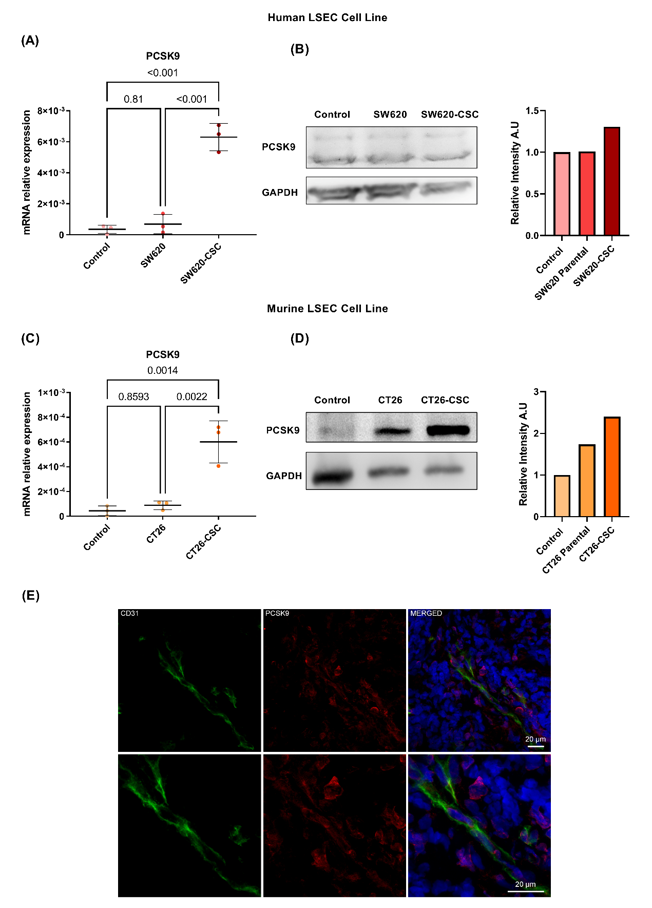
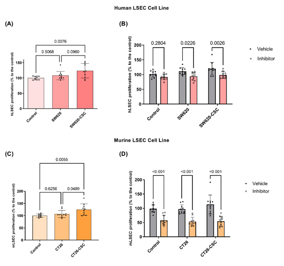
Inhibiting PCSK9 Attenuates Liver Endothelial Cell Activation During Liver Metastasis
Colorectal cancer (CRC) often leads to liver metastases, a major cause of patient mortality. This study investigated the role of proprotein convertase subtilisin/kexin type 9 (PCSK9), a protein lately related to the metastatic process, focusing on its effects on liver sinusoidal endothelial cells (LSECs), key components of the liver microenvironment.
LSECs were stimulated with conditioned media derived from differentiated colorectal cancer cells and cancer stem cells (CSCs). RNA sequencing was used to profile gene expression in LSECs. PCSK9 mRNA and protein levels were quantified by qPCR and Western blotting, respectively. PCSK9 expression in CRC liver metastases was evaluated by immunofluorescent staining.
Researchers found that conditioned media from colorectal cancer stem cells strongly upregulates PCSK9 expression in LSECs. PCSK9 activation enhances LSEC proliferation and migration, contributing to the formation of a pro-metastatic niche. Inhibiting PCSK9 with the small molecule PF-06446864 reduced endothelial activation and normalized gene expression, decreasing LSEC potential support of the metastasis. Immunofluorescence confirmed PCSK9 expression in LSECs of human colorectal cancer liver metastases. These results suggest that PCSK9 could represent a promising therapeutic target to prevent and treat liver metastases in colorectal cancer patients.
Ask a Question
Write your own review

Description: Nasal epithelial cells form the outermost protective layer against environmental factors. They clean, humidify, and warm inhaled air. They produces mucus, which binds particles that are subsequently ...

Description: Immortalized Human Corneal Epithelial Cells-SV40 have been obtained immortalizing Human Corneal Epithelial Cells with Lenti-SV40 Lentivirus. Immortalized cells were controlled passaging side by side ...
Description: Immortalized Human Lymphatic Endothelial Cells-SV40 were developed from human tissues transduced with a lentiviral expression vector containing the SV40T gene. The cell line was continuously cultured ...
Description: Immortalized Human Retinal Pigment Epithelial Cells were isolated from neonatal human globes and spontaneously immortalized in culture bypassing crisis. They retained typical morphology of the ...
- Adipose Tissue-Derived Stem Cells
- Human Neurons
- Mouse Probe
- Whole Chromosome Painting Probes
- Hepatic Cells
- Renal Cells
- In Vitro ADME Kits
- Tissue Microarray
- Tissue Blocks
- Tissue Sections
- FFPE Cell Pellet
- Probe
- Centromere Probes
- Telomere Probes
- Satellite Enumeration Probes
- Subtelomere Specific Probes
- Bacterial Probes
- ISH/FISH Probes
- Exosome Isolation Kit
- Human Adult Stem Cells
- Mouse Stem Cells
- iPSCs
- Mouse Embryonic Stem Cells
- iPSC Differentiation Kits
- Mesenchymal Stem Cells
- Immortalized Human Cells
- Immortalized Murine Cells
- Cell Immortalization Kit
- Adipose Cells
- Cardiac Cells
- Dermal Cells
- Epidermal Cells
- Peripheral Blood Mononuclear Cells
- Umbilical Cord Cells
- Monkey Primary Cells
- Mouse Primary Cells
- Breast Tumor Cells
- Colorectal Tumor Cells
- Esophageal Tumor Cells
- Lung Tumor Cells
- Leukemia/Lymphoma/Myeloma Cells
- Ovarian Tumor Cells
- Pancreatic Tumor Cells
- Mouse Tumor Cells