
Immortalized Human Gingival Fibroblasts-hTERT
Cat.No.: CSC-I9082L
Species: Homo sapiens
Source: Gingiva
Morphology: spindle-shaped
Culture Properties: Adherent
- Specification
- Background
- Scientific Data
- Q & A
- Customer Review
Note: Never can cells be kept at -20 °C.
CIK-HT013 HT® Lenti-hTERT Immortalization Kit
2. MTT assay was used to confirm the proliferative potential of the immortalized cells over the 70th passage.
Immortalized human gingival fibroblasts-hTERT (HGF/hTERT) are generated by ectopic expression of human telomerase reverse transcriptase in primary human gingival fibroblasts. Unlike SV40 large T antigen, hTERT extends telomeres without directly inactivating p53 or Rb, thereby preserving more normal cell-cycle checkpoints, stress responses, and genomic stability. This yields an extended lifespan while maintaining core fibroblast features: vimentin, S100A4/FSP1, CD90/THY1, collagen I/III, fibronectin, and TGF-β–inducible α-smooth muscle actin.
Functionally, HGF/hTERT retain extracellular matrix synthesis and remodeling, wound-healing migration, and responsiveness to inflammatory cytokines and microbial ligands through Toll-like receptor signaling. They are therefore well suited to study gingival homeostasis, periodontal inflammation, fibrosis, gingival overgrowth, oral mucositis, and host–microbiome interactions. They also support biocompatibility and toxicity testing of dental materials, oral care products, and pharmaceutical compounds.
Compared with primary HGFs, HGF/hTERT provide a continuous, scalable, and cost-effective source with high batch-to-batch consistency and reduced donor variability. This enables reproducible high-throughput screening, long-term culture, siRNA/CRISPR perturbation, and co-culture with gingival epithelial cells or immune cells.
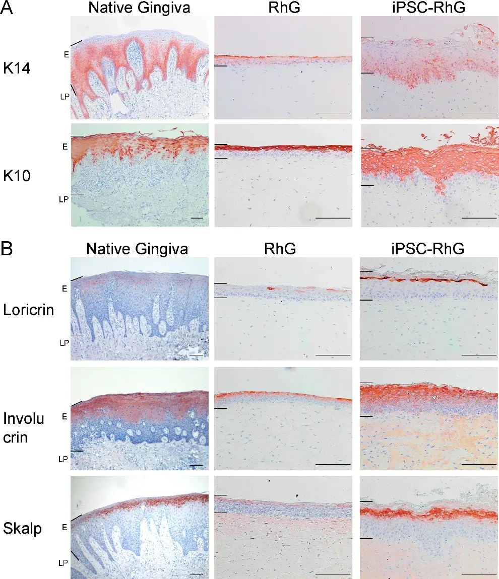
Incorporation of iPSCs Together with TERT-Immortalized Keratinocytes and Fibroblasts into Reconstructed Human Gingiva Enhances Phenotype of Gingival Epithelium
Current reconstructed human gingiva models (RhG) serve as valuable testing platforms for safety and efficacy testing of dental materials, however they lack important phenotypic characteristics typical of the gingival epithelium. The aim of this study was to determine whether incorporating iPSCs into the hydrogel of the current cell line RhG would improve its phenotype.
Immortalized human gingival fibroblasts were resuspended with and without iPSCs in collagen-fibrin hydrogels and gingival keratinocytes were seeded on top of the hydrogels to construct RhGs. RhGs were cultured at air-liquid interface for 1, 2, 4 and 6 weeks and extensively characterized by immunohistochemistry. In situ hybridization for X and Y chromosomes was conducted to identify female iPSCs and male fibroblasts in the RhGs.
Compared to the standard RhG without iPSCs, models containing a mix of fibroblasts and iPSCs in the hydrogel (iPSC-RhGs) showed an increased proliferation of the keratinocytes within the epithelium, increased epithelial thickness, improved differentiation and even invagination of the epithelium into the collagen hydrogel, thus resulting in an epithelial phenotype much more resembling that of the native gingiva.
Ask a Question
Write your own review
Description: Immortalized Human Gastric Epithelial Cells-SV40 have been obtained immortalizing Human Gastric Epithelial Cells with Lenti-SV40 Lentivirus. Immortalized cells were controlled passaging side by side ...
Description: Immortalized Human Kidney Podocytes-SV40 have been obtained immortalizing Human Kidney Podocytes with Lenti-SV40 Lentivirus. Immortalized cells were controlled passaging side by side with the primary ...
Description: Nasal epithelial cells form the outermost protective layer against environmental factors. They clean, humidify, and warm inhaled air. They produces mucus, which binds particles that are subsequently ...
Description: Immortalized Human Splenic Endothelial Cells-SV40 have been obtained immortalizing Human Splenic Endothelial Cells with SV40LT expressing lentiviral particles. Immortalized cells were controlled ...
Description: Immortalized Human Corneal Epithelial Cells-SV40 have been obtained immortalizing Human Corneal Epithelial Cells with Lenti-SV40 Lentivirus. Immortalized cells were controlled passaging side by side ...
- Adipose Tissue-Derived Stem Cells
- Human Neurons
- Mouse Probe
- Whole Chromosome Painting Probes
- Hepatic Cells
- Renal Cells
- In Vitro ADME Kits
- Tissue Microarray
- Tissue Blocks
- Tissue Sections
- FFPE Cell Pellet
- Probe
- Centromere Probes
- Telomere Probes
- Satellite Enumeration Probes
- Subtelomere Specific Probes
- Bacterial Probes
- ISH/FISH Probes
- Exosome Isolation Kit
- Human Adult Stem Cells
- Mouse Stem Cells
- iPSCs
- Mouse Embryonic Stem Cells
- iPSC Differentiation Kits
- Mesenchymal Stem Cells
- Immortalized Human Cells
- Immortalized Murine Cells
- Cell Immortalization Kit
- Adipose Cells
- Cardiac Cells
- Dermal Cells
- Epidermal Cells
- Peripheral Blood Mononuclear Cells
- Umbilical Cord Cells
- Monkey Primary Cells
- Mouse Primary Cells
- Breast Tumor Cells
- Colorectal Tumor Cells
- Esophageal Tumor Cells
- Lung Tumor Cells
- Leukemia/Lymphoma/Myeloma Cells
- Ovarian Tumor Cells
- Pancreatic Tumor Cells
- Mouse Tumor Cells