
HLC-1
Cat.No.: CSC-C6528J
Species: Homo sapiens (Human)
Source: Pleural Effusion Metastasis
Morphology: epithelial-like
Culture Properties: Adherent cells
- Specification
- Background
- Scientific Data
- Q & A
- Customer Review
Store in liquid nitrogen.
HLC-1 (Human lung carcinoma-1) is a cell line derived from primary lung carcinoma of a human patient. It is frequently described as a non-small cell lung cancer (NSCLC) cell line model. HLC-1 has been used extensively as a model of respiratory disease and lung cancer because of its stability and epithelial characteristics.
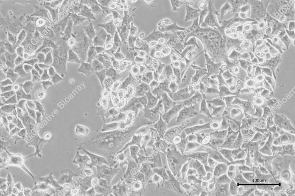
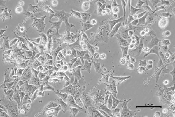
The HLC-1 cell line displays epithelial-like morphology and growth as an adherent monolayer. They are polygonal to cobblestone in appearance, as is typical of lung carcinoma cells. HLC-1 cells were shown to express epithelial markers. Furthermore, expression and regulation of proteins involved in cellular proliferation, cell survival, and differentiation are dysregulated. An analysis of the karyotype from HLC-1 cells identified chromosomal changes associated with malignant transformation of lung tumors. Researchers employ this cell line to investigate dysregulated cell cycle progression along with apoptosis evasion mechanisms as well as responses to environmental and chemical insults during lung carcinogenesis. HLC-1 cells have been used to assess the cytotoxicity and effects of various anticancer drugs, air pollutants, and inhaled chemicals on lung epithelial cells.
Green Synthesized Ag Nanoparticles Decorated on the Surface of Hibiscus Tea Leaf Extract Modified Reduced Graphene Oxide: Investigation of its Anti-Lung Carcinoma Effects
Green synthesis of silver nanoparticles on reduced graphene oxide (RGO/Ag NPs) using Hibiscus tea leaf extract is eco-friendly and yields uniform, spherical particles. Wu's team evaluated the anticancer potential of this nanocomposite against lung carcinoma cells and its antioxidant activity.
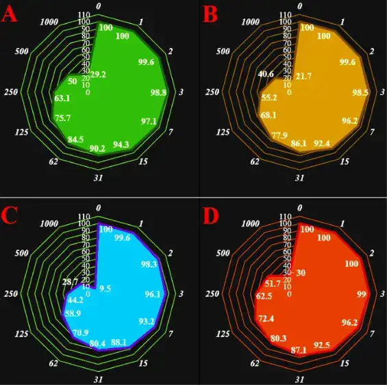
The Hibiscus-assisted graphene nanocomposite dose dependently decreased the viability of human lung carcinoma cell lines HLC-1, PC-14, LC-2/ad and NCI-H322 with IC50 values of 500, 327, 204 and 542 µg/mL, respectively (Fig. 1). In another similar work Alsaedi et al. (2019) prepared reduced graphene oxide (RGO) through ultrasonic processing and studied the cytotoxicity of prepared RGO against MCF-7 breast carcinoma cells using comet assay, AO-EB staining ROS formation, mitochondrial membrane potential, NF-κB translocation along with MTT assay. Expression studies of Bax and Bcl-2 using qPCR and fluorescence microscopy showed that RGO only mediated apoptosis through mitochondrial signaling pathway along with involvement of NF-κB pathway.
Ask a Question
Write your own review
- You May Also Need

Description: Lung small cell carcinoma producing insulin-like growth factor II. Cell growth is slow.

Description: Lung Cancer-1/squamous. Parent cell line of LC-1/sq-SF, the same patient as LC-F. Cell growth is slow.

Description: Species: human - male, 47 years old, CaucasianTumorigenecity: does not produce tumorsIsoenzyme: Me-2, 1-2; PGM3, 1-2; PGM1, 1; ES D, 2; AK1, 1; GLO-1, 1-2; G6PD, BHistopathology: carcinoma, ...

Description: Established in 1979 from the bone marrow aspirated from a 55-year-old white man with small cell lung carcinoma prior to treatment; corresponds to NCI-H209; described as expressing neuroendocrine ...

- Adipose Tissue-Derived Stem Cells
- Human Neurons
- Mouse Probe
- Whole Chromosome Painting Probes
- Hepatic Cells
- Renal Cells
- In Vitro ADME Kits
- Tissue Microarray
- Tissue Blocks
- Tissue Sections
- FFPE Cell Pellet
- Probe
- Centromere Probes
- Telomere Probes
- Satellite Enumeration Probes
- Subtelomere Specific Probes
- Bacterial Probes
- ISH/FISH Probes
- Exosome Isolation Kit
- Human Adult Stem Cells
- Mouse Stem Cells
- iPSCs
- Mouse Embryonic Stem Cells
- iPSC Differentiation Kits
- Mesenchymal Stem Cells
- Immortalized Human Cells
- Immortalized Murine Cells
- Cell Immortalization Kit
- Adipose Cells
- Cardiac Cells
- Dermal Cells
- Epidermal Cells
- Peripheral Blood Mononuclear Cells
- Umbilical Cord Cells
- Monkey Primary Cells
- Mouse Primary Cells
- Breast Tumor Cells
- Colorectal Tumor Cells
- Esophageal Tumor Cells
- Lung Tumor Cells
- Leukemia/Lymphoma/Myeloma Cells
- Ovarian Tumor Cells
- Pancreatic Tumor Cells
- Mouse Tumor Cells