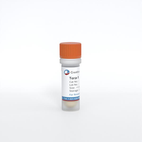
Tera-1
Cat.No.: CSC-C9736L
Species: Homo sapiens (Human)
Source: Lung Metastasis
Morphology: epithelial
Culture Properties: monolayer
- Specification
- Background
- Scientific Data
- Q & A
- Customer Review
Tumorigenecity: does not produce tumors
Isoenzyme: Me-2, 1-2; PGM3, 1-2; PGM1, 1; ES D, 2; AK1, 1; GLO-1, 1-2; G6PD, B
Histopathology: carcinoma, embryonal; metastasis to lung
vWA: 14
FGA: 21,25
Amelogenin: X
TH01: 7,8
TPOX: 8
CSF1P0: 11
D5S818: 11,13
D13S317: 12
D7S820: 9,10
Shipping Condition: Room Temperature
Tera-1 is an immortalized human embryonal carcinoma (EC) cell line. Tera-1 was originally established from testicular tumor tissue from a 26-year-old man with localized, advanced gonadal germ cell tumor (GCT) in 1980. As a subtype of non-seminomatous GCTs, the parental cells of Tera-1 cell line are derived from primordial germ cells in the seminiferous tubules of the testes, sharing the essential pathological properties of high-grade embryonal carcinoma. Molecularly, the Tera-1 cells highly express pluripotent stem cell markers (Oct4, Nanog, Sox2) and EC-specific makers (PLAP, CD30), and activate the signaling pathways related to embryonic development (Wnt/β-catenin, PI3K-AKT).
Functionally, it maintains multi-lineage differentiation ability (able to differentiate into endoderm, mesoderm and ectoderm cells when induced by retinoic acid) and malignant phenotype (invaded in Transwell assays, 100% subcutaneous tumor formation in nude mice). Tera-1 cell line is widely used as a model to study the pathogenesis of GCTs (such as differentiation arrest of germ cells), regulation of embryonic development, screening of anti-GCT drugs (such as cisplatin sensitivity assay), reproductive toxicants evaluation, and exploration of germ cell protection methods.
Proteins and Parameters of HML.2 from Teratocarcinoma Cell Lines GH and Tera-1
Approximately 8% of our genome consists of sequences from Human Endogenous Retroviruses (HERVs). The HERV-K (HML.2) family, abbreviated here as HML.2, produces virus particles detected in cell lines, malignant tumors, and autoimmune diseases. Morozov et al. aim to characterize the virus proteins, estimate the buoyant density of viruses, and measure the RT activity of HML.2 GH and HML.2 Tera-1.
In order to facilitate the isolation of the viruses, GH and Tera-1 cells were cultivated in the normal medium without addition of any hormones or other stimulants. The supernatants of the cells were collected and processed the same day to prevent destruction of the virions. The virus pellets were diluted 2000 times and used for analysis in Western blot with anti-p27-CA serum (Fig. 1a). GH cells produced 4-5 times more Gag proteins compared to Tera-1 cells. HML.2 GH contained more unprocessed Pr76Gag, which may indicate impaired capsid maturation and Tera-1 had less unprocessed Gag precursors. Intermediate cleavage products of 50-42 kDa were present in both preparations, suggesting the presence of some virions with incompletely assembled capsids (Fig. 1a). To estimate viral buoyant density and to analyze the protein distribution, they performed sucrose density gradient centrifugation (Dgc). After Dgc, the fractions were collected, diluted in TN buffer, pelleted and used for analysis in Western blot (Fig. 1b, c). The majority of CA and TM proteins were found in fraction 4 (F4) with a buoyant density of 1.16 g/cm³. "Light" content, mainly Pr76Gag, was found in F3 (1.135 g/cm³) (Fig. 1b) which may be due to interaction with extracellular vesicles or low vRNA content.

VNC Drug Internalization of Tera-1 Cells
Vincristine (VNC), a powerful anticancer drug, faces challenges such as cytotoxicity and nonspecific distribution, leading to severe side effects. To address these issues, Al-Musawi et al. have explored using superparamagnetic iron oxide nanoparticles (SPIONs) encapsulated in dextran and conjugated with folate to deliver VNC specifically to cancer cells. They investigated the anticancer activity and molecular mechanisms of apoptosis induced by this novel polymeric-magnetic nanocarrier in Tera-1 testicular tumor cells.
In Tera-1 cancer cells, the introduced FA-DEX-VNC-SPION system was successfully internalized and it is clearly observed by fluorescence microscopy (Fig. 2B, C), while VNC was present in the form of crystal bodies of different sizes (Fig. 2A). The cells were treated with VNC-loaded nanocarriers and they were bright green because of the high internalization capacity. This finding was achieved due to the increasing solubility level of VNC after loading the nanocarrier. Green, star-like, and insoluble fragments were noticed in cells treated with VCN alone due to their insolubility in the aqueous medium. This finding clearly reveals that FA-DEX-VNC-SPION is a really powerful carrier which can be used as a delivery mechanism for drugs in Tera-1 cancer cell-targeted therapy. The results were considered satisfactory in the target applications and are in accordance with those previously reported.

Ask a Question
Write your own review
- You May Also Need
- Adipose Tissue-Derived Stem Cells
- Human Neurons
- Mouse Probe
- Whole Chromosome Painting Probes
- Hepatic Cells
- Renal Cells
- In Vitro ADME Kits
- Tissue Microarray
- Tissue Blocks
- Tissue Sections
- FFPE Cell Pellet
- Probe
- Centromere Probes
- Telomere Probes
- Satellite Enumeration Probes
- Subtelomere Specific Probes
- Bacterial Probes
- ISH/FISH Probes
- Exosome Isolation Kit
- Human Adult Stem Cells
- Mouse Stem Cells
- iPSCs
- Mouse Embryonic Stem Cells
- iPSC Differentiation Kits
- Mesenchymal Stem Cells
- Immortalized Human Cells
- Immortalized Murine Cells
- Cell Immortalization Kit
- Adipose Cells
- Cardiac Cells
- Dermal Cells
- Epidermal Cells
- Peripheral Blood Mononuclear Cells
- Umbilical Cord Cells
- Monkey Primary Cells
- Mouse Primary Cells
- Breast Tumor Cells
- Colorectal Tumor Cells
- Esophageal Tumor Cells
- Lung Tumor Cells
- Leukemia/Lymphoma/Myeloma Cells
- Ovarian Tumor Cells
- Pancreatic Tumor Cells
- Mouse Tumor Cells
