RI-1
Cat.No.: CSC-C0590
Species: Homo sapiens (Human)
Source: Blood; Peripheral Blood
Morphology: large round cells growing in suspension
Culture Properties: suspension
- Specification
- Background
- Scientific Data
- Q & A
- Customer Review
Immunology: CD3 -, CD10 -, CD13 -, CD20 +, CD34 -, CD37 +, CD38 +, CD80 +, HLA-DR +, sm/cyIgG -, sm/cyIgM +, sm/cykappa +, sm/cylambda -
Viruses: PCR: EBV -, HBV -, HCV -, HIV -, HTLV-I/II -, SMR
RI-1 is a human B-cell non-Hodgkin lymphoma (B-NHL) cell line, originally established from a patient with diffuse large B-cell lymphoma (DLBCL). This cell line is typically used for in vitro research and is known for its stable growth in suspension culture. Phenotypically, RI-1 cells are positive for B-cell markers such as CD19, CD20, CD22, and surface immunoglobulin, consistent with mature B-cell lineage. At the molecular level, RI-1 cells exhibit alterations in several signaling pathways commonly implicated in B-cell lymphomas, including B-cell receptor (BCR) signaling, NF-κB activation, and apoptosis regulation. Genetically, RI-1 cells possess genetic lesions often associated with B-cell lymphomas, providing a relevant model for the study of B-cell lymphoma pathogenesis and treatment.
RI-1 cell line is commonly used for studying B-cell lymphoma pathogenesis and signal transduction, as well as for preclinical testing of immune-targeted therapies. This cell line is also valuable for the screening and evaluation of potential therapeutic agents targeting B-cell surface antigens, BCR signaling components, and apoptosis regulators, supporting lymphoma research and drug discovery efforts.
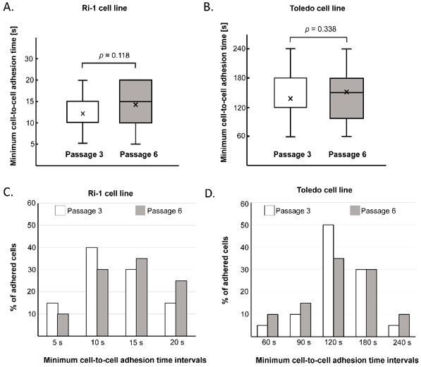
Determination of the Minimum Cell-to-Cell Adhesion Time Using Optical Tweezers in Leukemia and Lymphoma Research
Single-cell adhesion assays study attachment and detachment. Optical tweezers (OTs) are great for this because they're gentle and don't need labels. OTs can measure tiny forces and study how long cells need to touch to stick together.
Duś-Szachniewicz et al. made a step-by-step guide to use OTs with low laser power to study how leukemia-lymphoma cells stick to other cells. Their method is very precise and can spot small changes in cell adhesion, as shown in Figure 1. They found big differences between two cell lines, Ri-1 and Toledo. Ri-1 cells stuck to other cells in 5-20 seconds, while Toledo cells took 60-240 seconds. The average time for Ri-1 was 12.83 ± 4.94 seconds, much faster than Toledo's 141 ± 49.69 seconds. Figures 1C and D show how many cells stuck over time. They tested both early (passage 3) and later (passage 6) cells and found no big differences. Their results match a previous study and suggest that different cell lines have different surface properties or adhesion mechanisms. Optical tweezers are way better than traditional methods, which can only measure adhesion times of 30-60 minutes and often miss important details. Single-cell studies help us understand cell differences and how tissues and organs work.
Ask a Question
Write your own review
- You May Also Need

Description: Established in 2007 from the bone marrow mononuclear cells of an 82-year-old Japanese man with diffuse large B-cell lymphoma in the leukemic phase

Description: Established from the bone marrow of a 28-year-old man who developed the terminal leukemic phase of lymphosarcoma in 1976

Description: This cell line was derived from the bone marrow aspirate of a 59 year old male with erythroleukemia that became acute myelogenous leukaemia.The cells form colonies in soft-agar in the presence of ...

Description: Established from the pleural effusion of a 24-year-old woman with recurrent anaplastic large cell lymphoma (ALCL); cells were described to clonally derive from T-lineage lymphoid cells and to be ...

Description: Established from a 37-year-old man at second (refractory/terminal) relapse of Hodgkin lymphoma (nodular sclerosing -> lymphocyte depleted/stage IIISA -> stage IV) after both combined chemo- and ...

Description: Established from the peripheral blood of a 10-year-old Caucasian boy with acute lymphoblastic leukemia (pre B-ALL) at diagnosis in 1993
- Adipose Tissue-Derived Stem Cells
- Human Neurons
- Mouse Probe
- Whole Chromosome Painting Probes
- Hepatic Cells
- Renal Cells
- In Vitro ADME Kits
- Tissue Microarray
- Tissue Blocks
- Tissue Sections
- FFPE Cell Pellet
- Probe
- Centromere Probes
- Telomere Probes
- Satellite Enumeration Probes
- Subtelomere Specific Probes
- Bacterial Probes
- ISH/FISH Probes
- Exosome Isolation Kit
- Human Adult Stem Cells
- Mouse Stem Cells
- iPSCs
- Mouse Embryonic Stem Cells
- iPSC Differentiation Kits
- Mesenchymal Stem Cells
- Immortalized Human Cells
- Immortalized Murine Cells
- Cell Immortalization Kit
- Adipose Cells
- Cardiac Cells
- Dermal Cells
- Epidermal Cells
- Peripheral Blood Mononuclear Cells
- Umbilical Cord Cells
- Monkey Primary Cells
- Mouse Primary Cells
- Breast Tumor Cells
- Colorectal Tumor Cells
- Esophageal Tumor Cells
- Lung Tumor Cells
- Leukemia/Lymphoma/Myeloma Cells
- Ovarian Tumor Cells
- Pancreatic Tumor Cells
- Mouse Tumor Cells