
P30-OHKUBO
Cat.No.: CSC-C6233X
Species: Homo sapiens (Human)
Source: Bone Marrow
Morphology: single cells growing in suspension
Culture Properties: suspension
- Specification
- Background
- Scientific Data
- Q & A
- Customer Review
Immunology: CD3 -, CD10 +, CD13 -, CD19 +, CD20 +, CD34 -, CD37 -, CD38 +, cyCD79a +, CD80 -, CD138 +, HLA-DR +, cyIgG -, cyIgM +, cykappa -, cylambda +
Viruses: PCR: EBV -, HBV -, HCV -, HIV -,
P30-OHKUBO is a human pancreatic cancer cell line derived from pancreatic ductal adenocarcinoma (PDAC). It is commonly used as an in vitro model system to study the biology and molecular mechanisms of pancreatic cancer. This aggressive cancer is characterized by rapid growth, invasion, and resistance to treatment. P30-OHKUBO cells are epithelial in morphology and are typically cultured as adherent cells.
This cell line possesses genetic and phenotypic characteristics that are similar to pancreatic ductal adenocarcinoma including alterations in common oncogenic drivers and signaling pathways involved in PDAC such as KRAS and downstream effectors that control cell proliferation, survival, and invasion. The P30-OHKUBO cell line can be used to study tumor growth, metastasis, and drug resistance.
Common uses for this cell line include proliferation assays, migration and invasion assays, apoptosis assays, and drug sensitivity assays. Additionally, P30-OHKUBO can be used in xenograft models to assess tumorigenicity and response to therapeutics in vivo. Overall, P30-OHKUBO can be used as an experimental model system for mechanistic studies and preclinical studies of pancreatic cancer.
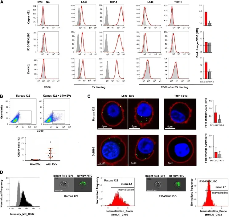
CD30+ EVs Enable the Targeting of CD30- DLBCL Cells by the CD30 Antibody-Drug Conjugate Brentuximab Vedotin
Brentuximab Vedotin (BV), an anti-CD30 antibody-drug conjugate, shows efficacy in CD30-negative diffuse large B-cell lymphoma (DLBCL), but the mechanism is unclear. Lobastova et al. investigated whether CD30+ extracellular vesicles (EVs) enable BV binding and uptake in CD30-negative tumor cells.
To study CD30+ EVs mediating BV toxicity in non-Hodgkin B cell leukemia, they assessed three target cell lines for endogenous CD30, EV binding, and CD30 enrichment. Flow cytometry showed Karpas 422 and DoHH-2 lacked CD30, while P30-OH/KUBO was positive (Fig. 1A, left). All lines bound fluorescent EVs from L540 and THP-1 cells (Fig. 1A, middle). Only L540-derived EVs increased CD30 on CD30- cell surfaces (Fig. 1A, right). CD30 enrichment was measured by comparing CD30 MFI in EV-treated vs. untreated cells. Karpas 422 showed better CD30 surface enrichment than DoHH-2 after 2 h; in CD30+ P30-OH/KUBO, EVs contributed little to total CD30. Membrane-labeled EVs gave stronger signals than anti-CD30 staining. Only a small fraction of target cells bound CD30+ EVs (Fig. 1B), likely due to EV heterogeneity. CD30 internalization may also differ by cell type. We then studied anti-CD30 internalization. After co-incubating cells with EVs and fluorescent anti-CD30 (using SGN30 or Ki-3 due to BV labeling issues), confocal microscopy revealed CD30+ EVs promoted antibody uptake in Karpas 422 and DoHH-2, but not with THP-1 EVs (Fig. 1C). Intracellular signals exceeded surface signals, partly explaining low surface CD30 in prior assays. Imaging flow cytometry quantified SGN30-FITC uptake, yielding internalization scores of 3.1 (Karpas 422, N=2156) and 2.1 (P30-OH/KUBO, N=1086) with L540 EVs, confirming CD30+ EVs enable antibody uptake (Fig. 1D).
Ask a Question
Write your own review
- You May Also Need

Description: Established in 2007 from the bone marrow mononuclear cells of an 82-year-old Japanese man with diffuse large B-cell lymphoma in the leukemic phase

Description: Established from the bone marrow of a 28-year-old man who developed the terminal leukemic phase of lymphosarcoma in 1976

Description: This cell line was derived from the bone marrow aspirate of a 59 year old male with erythroleukemia that became acute myelogenous leukaemia.The cells form colonies in soft-agar in the presence of ...

Description: Established from the pleural effusion of a 24-year-old woman with recurrent anaplastic large cell lymphoma (ALCL); cells were described to clonally derive from T-lineage lymphoid cells and to be ...

Description: Established from a 37-year-old man at second (refractory/terminal) relapse of Hodgkin lymphoma (nodular sclerosing -> lymphocyte depleted/stage IIISA -> stage IV) after both combined chemo- and ...

Description: Established from the peripheral blood of a 10-year-old Caucasian boy with acute lymphoblastic leukemia (pre B-ALL) at diagnosis in 1993
- Adipose Tissue-Derived Stem Cells
- Human Neurons
- Mouse Probe
- Whole Chromosome Painting Probes
- Hepatic Cells
- Renal Cells
- In Vitro ADME Kits
- Tissue Microarray
- Tissue Blocks
- Tissue Sections
- FFPE Cell Pellet
- Probe
- Centromere Probes
- Telomere Probes
- Satellite Enumeration Probes
- Subtelomere Specific Probes
- Bacterial Probes
- ISH/FISH Probes
- Exosome Isolation Kit
- Human Adult Stem Cells
- Mouse Stem Cells
- iPSCs
- Mouse Embryonic Stem Cells
- iPSC Differentiation Kits
- Mesenchymal Stem Cells
- Immortalized Human Cells
- Immortalized Murine Cells
- Cell Immortalization Kit
- Adipose Cells
- Cardiac Cells
- Dermal Cells
- Epidermal Cells
- Peripheral Blood Mononuclear Cells
- Umbilical Cord Cells
- Monkey Primary Cells
- Mouse Primary Cells
- Breast Tumor Cells
- Colorectal Tumor Cells
- Esophageal Tumor Cells
- Lung Tumor Cells
- Leukemia/Lymphoma/Myeloma Cells
- Ovarian Tumor Cells
- Pancreatic Tumor Cells
- Mouse Tumor Cells