KE-37
Cat.No.: CSC-C0218
Species: Homo sapiens (Human)
Morphology: single, round cells in suspension
Culture Properties: suspension
- Specification
- Background
- Scientific Data
- Q & A
- Customer Review
Immunology: CD2 +, cyCD3 +, smCD3 -, CD4 +, CD5 +, CD6 +, CD7 +, CD8 +, CD13 -, CD19 -, CD34 -, TCRalpha/b
KE-37 is a human cell line derived from peripheral blood of a 27-year-old man in 1979. The human cell line is used as a model system in the study of immunology and hematology. The cell line is known as a T-cell acute lymphoblastic leukemia (T-ALL) cell line. KE-37 has been used as a model to study the molecular pathogenesis of T-cell malignancies and to test the efficacy of new chemotherapeutic agents and immunotherapies.
The KE-37 cell line demonstrates the characteristic of growing in suspension culture conditions. The cells have a round morphology and grow as single cells or small clusters. The cells show an immunophenotype of an immature or intermediate stage T-cell. The cell line is usually positive for CD2, CD4, and CD7 and negative for the mature T-cell receptor complex (CD3-). The cell line often contains a complex karyotype, including the most frequent translocation, t(8;14)(q24;q11), resulting in juxtaposition of the MYC oncogene to the T-cell receptor alpha/delta locus, seen in many cases of T-ALL.
KE-37 has also been at the center of several cell line authentication reports over the past few years. The recently reported widely used cell line SKW-3 has been found to be a derivative of KE-37 as a result of historic cross-contamination. KE-37 is used for high-throughput drug screening, signaling pathway studies (Notch1 or PI3K/AKT for example) and the development of CAR-T and bispecific antibody therapies for T-cell leukemias.
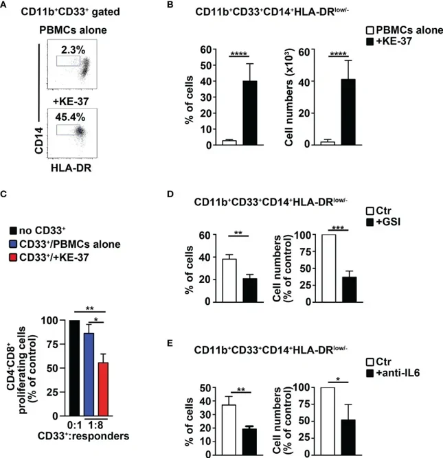
Human Notch-Dependent T-ALL Cell Lines Induce MDSCs From Healthy PBMCs
Notch receptors influence T-cell development, and their dysregulation is linked to T-ALL. MDSCs inhibit immune responses in tumors but are understudied in T-ALL. Here, Grazioli et al. used a Notch3-transgenic murine model of T-ALL to show that Notch-signaling deregulation promotes CD11b+Gr-1+ MDSCs.
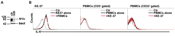
They extended their research to humans by coculturing PBMCs from healthy donors with the human Notch-dependent T-ALL cell line KE-37, which has Notch1 oncogenic mutations and is GSI resistant and Notch3 negative. KE-37 cells also express intracellular IL-6. In PBMCs/KE-37 cocultures, there was a significant increase in CD14+HLA-DRlow/neg MDSCs, both in percentage and absolute numbers, compared to PBMCs cultured alone (Fig. 1A, B). The CD33+ cells from these cocultures showed high suppressive function on autologous CD4-CD8+ T cells (Fig. 1C). KE-37 cells, which lack PTEN expression, do not respond to GSI treatment, although GSI blocks Notch1 activation. They confirmed Notch1 intracellular domain expression in KE-37 cells and its downregulation by GSI during cocultures (Fig. 2A). Repeating the coculture experiments with gamma-secretase inhibitors or neutralizing anti-IL-6 antibodies showed significant reductions in MDSC expansion (Fig. 1D, E), indicating that Notch activation and IL-6 signaling are crucial for MDSC expansion in these cocultures.
Ask a Question
Write your own review
- You May Also Need

Description: Established in 2007 from the bone marrow mononuclear cells of an 82-year-old Japanese man with diffuse large B-cell lymphoma in the leukemic phase

Description: Established from the bone marrow of a 28-year-old man who developed the terminal leukemic phase of lymphosarcoma in 1976

Description: This cell line was derived from the bone marrow aspirate of a 59 year old male with erythroleukemia that became acute myelogenous leukaemia.The cells form colonies in soft-agar in the presence of ...

Description: Established from the pleural effusion of a 24-year-old woman with recurrent anaplastic large cell lymphoma (ALCL); cells were described to clonally derive from T-lineage lymphoid cells and to be ...

Description: Established from a 37-year-old man at second (refractory/terminal) relapse of Hodgkin lymphoma (nodular sclerosing -> lymphocyte depleted/stage IIISA -> stage IV) after both combined chemo- and ...

Description: Established from the peripheral blood of a 10-year-old Caucasian boy with acute lymphoblastic leukemia (pre B-ALL) at diagnosis in 1993
- Adipose Tissue-Derived Stem Cells
- Human Neurons
- Mouse Probe
- Whole Chromosome Painting Probes
- Hepatic Cells
- Renal Cells
- In Vitro ADME Kits
- Tissue Microarray
- Tissue Blocks
- Tissue Sections
- FFPE Cell Pellet
- Probe
- Centromere Probes
- Telomere Probes
- Satellite Enumeration Probes
- Subtelomere Specific Probes
- Bacterial Probes
- ISH/FISH Probes
- Exosome Isolation Kit
- Human Adult Stem Cells
- Mouse Stem Cells
- iPSCs
- Mouse Embryonic Stem Cells
- iPSC Differentiation Kits
- Mesenchymal Stem Cells
- Immortalized Human Cells
- Immortalized Murine Cells
- Cell Immortalization Kit
- Adipose Cells
- Cardiac Cells
- Dermal Cells
- Epidermal Cells
- Peripheral Blood Mononuclear Cells
- Umbilical Cord Cells
- Monkey Primary Cells
- Mouse Primary Cells
- Breast Tumor Cells
- Colorectal Tumor Cells
- Esophageal Tumor Cells
- Lung Tumor Cells
- Leukemia/Lymphoma/Myeloma Cells
- Ovarian Tumor Cells
- Pancreatic Tumor Cells
- Mouse Tumor Cells