
- Specification
- Background
- Scientific Data
- Q & A
- Customer Review
FLO-1 is a continuous human esophageal adenocarcinoma (EAC) cell line derived from a primary distal esophageal adenocarcinoma, a tumor type frequently associated with Barrett’s esophagus. It exhibits epithelial morphology, adherent growth, and robust proliferation in standard culture, providing a reproducible and scalable in vitro model of EAC. FLO-1 cells express epithelial markers such as cytokeratins and E-cadherin, and are tumorigenic in immunodeficient mice, supporting subcutaneous and orthotopic xenograft studies.
Key advantages:
- Human EAC relevance: Represents distal esophageal adenocarcinoma, a cancer with rising incidence and poor prognosis, and is useful for studying Barrett’s-associated carcinogenesis and EAC biology.
- Experimental tractability: Amenable to transfection, siRNA/CRISPR perturbation, flow cytometry, immunostaining, qPCR, Western blot, and high-throughput drug screening.
- In vivo compatibility: Supports xenograft models for tumor growth, metastasis, and treatment response.
- Reproducibility: As a continuous line, it offers standardized culture, commercial availability, and reduced variability compared with primary tumors.
Applications: Chemoradiotherapy resistance, targeted therapy, invasion/metastasis, cancer stem cell biology, and biomarker discovery.
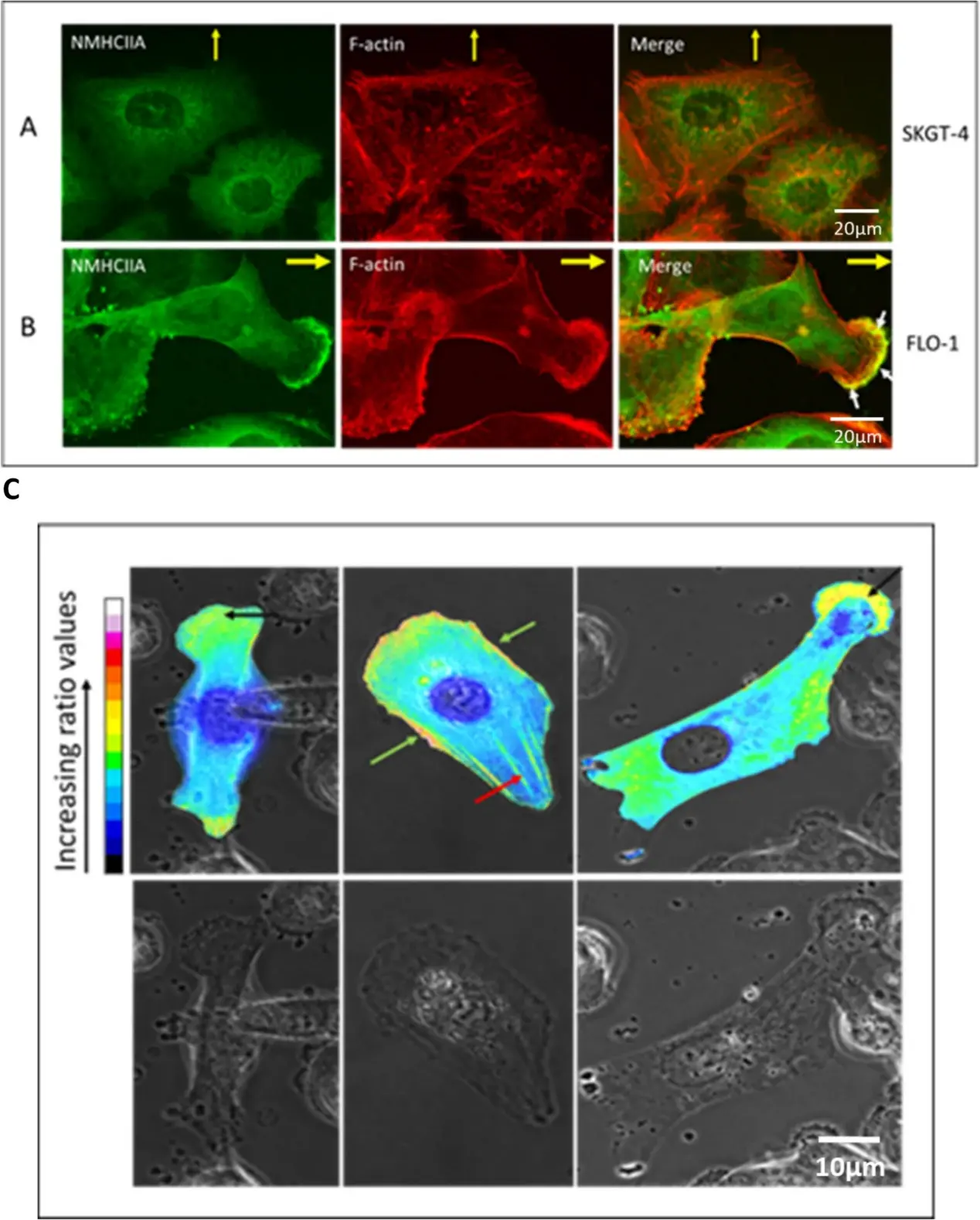
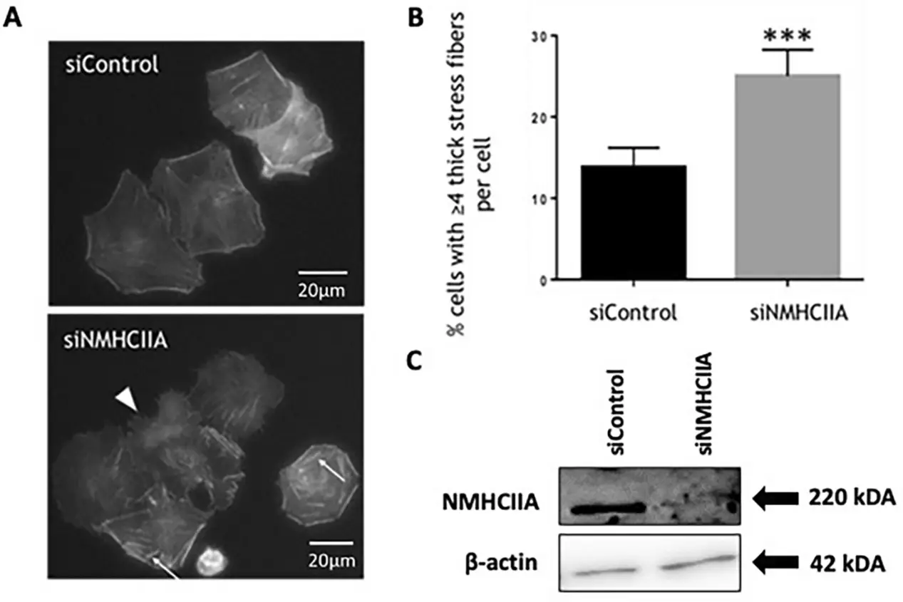
Non-Muscle Myosin Heavy Chain IIA Regulates Cell Morphology, Stress Fibre Structure, and Cell Migration in FLO-1 Oesophageal Adenocarcinoma Cells
Cell migration is tightly controlled in the healthy cell but can become dysregulated in diseases such as oesophageal adenocarcinoma (OAC) where increased cell motility and migration can contribute to metastasis. We investigated the role of an actin-based molecular motor, non-muscle myosin heavy chain IIA (NMHCIIA) in the migratory capacity of oesophageal adenocarcinoma cells.
Immunofluorescence microscopy and ratiometric imaging demonstrated that NMHCIIA co-localizes with F-actin at the leading edge and retracting rear of migrating FLO-1 OAC cells. siRNA-mediated depletion of NMHCIIA from FLO-1 cells altered cell morphology, gave rise to an increased number of stress fibre like structures and reduced FLO-1 cell migration. These findings suggest that NMHCIIA influences FLO-1 cell migration by regulating F-actin dynamics and the actin cytoskeleton, providing insight into the mechanisms of migration employed by OAC cells and identifying NMHCIIA as a potential therapeutic target for this disease.
Ask a Question
Write your own review
Description: OACP4 C was established from a primary tumour located in gastric cardia of a 55 year-old Caucasian male. The patient showed lymph node involvement with pleural metastases. OACP4 C was found to be ...

Description: Human poorly differentiated squamous cell carcinoma cell line established from esophageal cancer.

Description: Human moderately differentiated squamous cell carcinoma cell line established from esophageal cancer.

Description: Established from the primary tumor of a 73-year-old white woman with Barrett-esophageal adenocarcinoma in 1993

Description: Human moderately differentiated squamous cell carcinoma cell line established from esophageal cancer.

- Adipose Tissue-Derived Stem Cells
- Human Neurons
- Mouse Probe
- Whole Chromosome Painting Probes
- Hepatic Cells
- Renal Cells
- In Vitro ADME Kits
- Tissue Microarray
- Tissue Blocks
- Tissue Sections
- FFPE Cell Pellet
- Probe
- Centromere Probes
- Telomere Probes
- Satellite Enumeration Probes
- Subtelomere Specific Probes
- Bacterial Probes
- ISH/FISH Probes
- Exosome Isolation Kit
- Human Adult Stem Cells
- Mouse Stem Cells
- iPSCs
- Mouse Embryonic Stem Cells
- iPSC Differentiation Kits
- Mesenchymal Stem Cells
- Immortalized Human Cells
- Immortalized Murine Cells
- Cell Immortalization Kit
- Adipose Cells
- Cardiac Cells
- Dermal Cells
- Epidermal Cells
- Peripheral Blood Mononuclear Cells
- Umbilical Cord Cells
- Monkey Primary Cells
- Mouse Primary Cells
- Breast Tumor Cells
- Colorectal Tumor Cells
- Esophageal Tumor Cells
- Lung Tumor Cells
- Leukemia/Lymphoma/Myeloma Cells
- Ovarian Tumor Cells
- Pancreatic Tumor Cells
- Mouse Tumor Cells