
COLO-677
Cat.No.: CSC-C2560
Species: Homo sapiens (Human)
Source: Blood; Peripheral Blood
Morphology: mostly adherent round cells growing in monolayers
Culture Properties: monolayer
- Specification
- Background
- Scientific Data
- Q & A
- Customer Review
Immunology: cytokeratin -, cytokeratin-8 -, cytokeratin-18 -, desmin -, endothel -, GFAP -, HMB-45 -, neurofilament -, vimentin +
Viruses: ELISA: reverse transcriptase ne
COLO-677 cells were established from ascitic fluid of a patient with metastatic colorectal carcinoma. They exhibit morphology consistent with late passage colorectal cancer cells. These cells have been used primarily as an in vitro tumor model system for studying progression, therapy and chemoresistance mechanisms in colorectal cancer. As established cancer cells from ascites fluid, the cells grow as floating cells or loosely attached cells in culture, and rapidly proliferate in normal culture conditions.
The cells have been found to have a highly aneuploid karyotype. Dysregulated signaling in cell cycle arrest, apoptosis and metabolic changes have also been observed. Studies have utilized this cell line as a tool to understand tumor cell survival, oxidative stress and drug resistance in aggressive CRC. COLO-677 is also commonly used for drug screening or mechanistic studies to discover potential therapeutic targets.
Understanding the Radiobiological Mechanisms Induced by 177Lu-DOTATATE in Comparison to External Beam Radiation Therapy
177Lu-DOTATATE radionuclide therapy (RNT) effectively stabilizes neuroendocrine tumours (NETs), yet its radiobiology is largely borrowed from external-beam radiotherapy (EBRT). Delbart et al. compared key radiobiological endpoints and PARP-inhibitor (PARPi) radiosensitisation between 177Lu-DOTATATE and EBRT in SSTR-expressing cancer cells to determine whether RNT merits dedicated mechanistic studies.
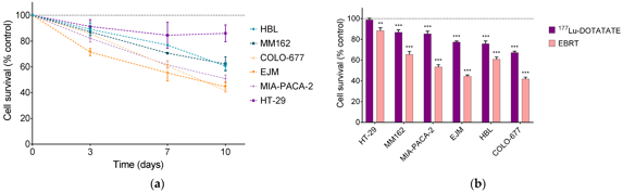
The radiosensitivity of six SSTR-expressing human cancer cell lines to ¹⁷⁷Lu-DOTATATE was previously characterized. Multiple myeloma lines (COLO-677: 33% ± 2%; EJM: 22% ± 2%) and melanoma HBL (26% ± 4%) showed highest sensitivity, with significant survival reduction at day 10. Intermediate sensitivity was observed in melanoma MM162 (13% ± 3%) and GEP MIA-PACA-2 (14% ± 3%), while GEP HT-29 was resistant. External beam radiotherapy (2 Gy) also induced time-dependent cytotoxicity (Fig. 1a), with day 10 survival of 42% (COLO-677), 45% (EJM), 51% (MIA-PACA-2), 61% (HBL), 62% (MM162), and 86% (HT-29)-the latter consistent with its reported radioresistance. EBRT produced significantly greater reduction than ¹⁷⁷Lu-DOTATATE in all lines. Notably, radiosensitivity rankings differed between modalities: HBL was as sensitive as myeloma lines to ¹⁷⁷Lu-DOTATATE but less responsive to EBRT (Fig. 1b).
Ask a Question
Write your own review
- You May Also Need

Description: Established in 2007 from the bone marrow mononuclear cells of an 82-year-old Japanese man with diffuse large B-cell lymphoma in the leukemic phase

Description: Established from the bone marrow of a 28-year-old man who developed the terminal leukemic phase of lymphosarcoma in 1976

Description: This cell line was derived from the bone marrow aspirate of a 59 year old male with erythroleukemia that became acute myelogenous leukaemia.The cells form colonies in soft-agar in the presence of ...

Description: Established from the pleural effusion of a 24-year-old woman with recurrent anaplastic large cell lymphoma (ALCL); cells were described to clonally derive from T-lineage lymphoid cells and to be ...

Description: Established from a 37-year-old man at second (refractory/terminal) relapse of Hodgkin lymphoma (nodular sclerosing -> lymphocyte depleted/stage IIISA -> stage IV) after both combined chemo- and ...

Description: Established from the peripheral blood of a 10-year-old Caucasian boy with acute lymphoblastic leukemia (pre B-ALL) at diagnosis in 1993
- Adipose Tissue-Derived Stem Cells
- Human Neurons
- Mouse Probe
- Whole Chromosome Painting Probes
- Hepatic Cells
- Renal Cells
- In Vitro ADME Kits
- Tissue Microarray
- Tissue Blocks
- Tissue Sections
- FFPE Cell Pellet
- Probe
- Centromere Probes
- Telomere Probes
- Satellite Enumeration Probes
- Subtelomere Specific Probes
- Bacterial Probes
- ISH/FISH Probes
- Exosome Isolation Kit
- Human Adult Stem Cells
- Mouse Stem Cells
- iPSCs
- Mouse Embryonic Stem Cells
- iPSC Differentiation Kits
- Mesenchymal Stem Cells
- Immortalized Human Cells
- Immortalized Murine Cells
- Cell Immortalization Kit
- Adipose Cells
- Cardiac Cells
- Dermal Cells
- Epidermal Cells
- Peripheral Blood Mononuclear Cells
- Umbilical Cord Cells
- Monkey Primary Cells
- Mouse Primary Cells
- Breast Tumor Cells
- Colorectal Tumor Cells
- Esophageal Tumor Cells
- Lung Tumor Cells
- Leukemia/Lymphoma/Myeloma Cells
- Ovarian Tumor Cells
- Pancreatic Tumor Cells
- Mouse Tumor Cells