
SCC-131
Cat.No.: CSC-C6667J
Species: Rattus norvegicus (Rat)
Morphology: Epithelial-Like
- Specification
- Background
- Scientific Data
- Q & A
- Customer Review
SCC‑131 is a human SCC cell line derived from an oral cavity tumor (squamous carcinoma of the floor of mouth). Originating from a tumor biopsy of a 73‑year‑old male, this cell line proliferates as adherent epithelial cells and features a typical doubling time of 50-60 hours when maintained under standard culture conditions.
The SCC‑131 cell line maintains many of the biological properties seen in tumor of origin, such as epithelial cell morphology and cytokeratin expression patterns consistent with squamous carcinomas. This cell line has been utilized as a model system for studying many molecular pathways relevant to oral cancer, such as response to chemotherapeutic agents like cisplatin and development of drug resistance. One study generated cisplatin‑resistant SCC131 /R sublines to study potential chemoresistance biomarkers that indicate cancer stem‑cell‑like and epithelial‑mesenchymal transition properties. Cells from this line can be used for various applications in basic and translational cancer research, such as studying tumor biology, drug screening, gene expression regulation, and the molecular basis of HNSCC.
QbD-Assisted Synthesis and Exploration of Plasmonic Laser Activatable Nanogold Seeds for Photothermal and Photodynamic Therapy of Cancer Cells
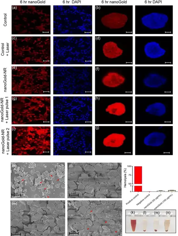
NanoGold seeds are promising for photothermal therapy due to their surface plasmon resonance properties. Gadeval et al. synthesized these seeds using a quality-by-design approach, targeting a 30-40 nm size range. To evaluate the anticancer potential of nanoGold seeds, cellular studies were conducted on both 2D monolayer and 3D spheroids models in SCC-131 cells. Solid tumor spheroids closely resemble the tumor environment in vivo as they are composed of proliferating cells at the outermost layer of tumor spheroid, quiescent cells at middle and necrotic cells at centre with gradient supply of nutrients and gases.
Cellular uptake studies compared nanoGold seeds-NR with plain Nile red solution. Results showed that Nile red alone did not enter cells significantly over 12 hours. NanoGold seeds-NR without NIR laser irradiation also showed minimal cellular uptake (Fig. 1e, f). However, nanoGold seeds-NR with NIR laser irradiation, especially dual-pulse irradiation (Fig. 1i, j), significantly enhanced cellular uptake compared to single-pulse irradiation (Fig. 1g, h). This suggests that NIR laser irradiation enhances the uptake of nanoGold seeds. The study also found that the cellular uptake of nanoGold seeds-NR improved over time, with significant red fluorescence intensity increases up to 6 hours, indicating that nanoGold seeds can enter and remain inside cells within 3-6 hours. This aligns with reports that NIR laser irradiation enhances the uptake of photothermally active nanocarriers, likely due to increased cell membrane mobility at elevated temperatures (42-45 °C).
Ask a Question
Write your own review
- You May Also Need
Description: Established from the pituitary tumor of an ovarectomized F344 rat treated with estrogen for 3 months; described as showing markedly reduced levels of TGF-β1 and TGF-β type II receptor mRNA; ...

Description: This cell line was derived from a 7-month-old female Wistar-Furth rat. GH3 cells produce growth hormone faster than the GH1 cell line and also produce prolactin. Hydrocortisone stimulates growth ...
Description: medullary thyroid carcinoma, neurotensin and calcitonin-producing, (WAG/RIJ rat)

Description: Ethyl nitrosourea induced tumour of the spinal cord and roots from a Wistar-Furth rat. In cell culture the cells grow to a high density and form a sheet of cells with many processes. TR33B is the ...
- Adipose Tissue-Derived Stem Cells
- Human Neurons
- Mouse Probe
- Whole Chromosome Painting Probes
- Hepatic Cells
- Renal Cells
- In Vitro ADME Kits
- Tissue Microarray
- Tissue Blocks
- Tissue Sections
- FFPE Cell Pellet
- Probe
- Centromere Probes
- Telomere Probes
- Satellite Enumeration Probes
- Subtelomere Specific Probes
- Bacterial Probes
- ISH/FISH Probes
- Exosome Isolation Kit
- Human Adult Stem Cells
- Mouse Stem Cells
- iPSCs
- Mouse Embryonic Stem Cells
- iPSC Differentiation Kits
- Mesenchymal Stem Cells
- Immortalized Human Cells
- Immortalized Murine Cells
- Cell Immortalization Kit
- Adipose Cells
- Cardiac Cells
- Dermal Cells
- Epidermal Cells
- Peripheral Blood Mononuclear Cells
- Umbilical Cord Cells
- Monkey Primary Cells
- Mouse Primary Cells
- Breast Tumor Cells
- Colorectal Tumor Cells
- Esophageal Tumor Cells
- Lung Tumor Cells
- Leukemia/Lymphoma/Myeloma Cells
- Ovarian Tumor Cells
- Pancreatic Tumor Cells
- Mouse Tumor Cells