NUGC-3
Cat.No.: CSC-C7088J
Species: Homo sapiens (Human)
Source: Muscle Metastasis
Morphology: Epithelial
Culture Properties: Adherent
- Specification
- Background
- Scientific Data
- Q & A
- Customer Review
The NUGC-3 cell line is a human-derived epithelial model established from a metastatic lymph node of a patient with gastric adenocarcinoma (GC). It represents a poorly differentiated (or diffuse-type) subtype according to the Lauren classification, which is often associated with aggressive behavior, early metastasis, and a less favorable prognosis compared to the intestinal-type. As a cell line derived from a metastatic site, NUGC-3 inherently models the advanced, disseminated stage of the disease, providing a critical tool for studying the mechanisms underlying gastric cancer progression and therapeutic resistance.
NUGC-3 cells exhibit an adherent, epithelial-like morphology in culture, though they may display a somewhat scattered growth pattern consistent with diffuse-type characteristics. A key molecular feature is its reported microsatellite instability-high (MSI-H) status, resulting from deficient DNA mismatch repair (dMMR). This genetic trait leads to a high tumor mutational burden (TMB), which has significant implications for the tumor's biological behavior and its potential response to immunotherapy. NUGC-3 is tumorigenic in immunocompromised mice, capable of forming tumors in vivo, and it generally retains a wild-type status for the TP53 gene, distinguishing it from many other GC models that harbor TP53 mutations.
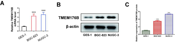
The Expression and Role of TMEM176B in Gastric Cancer Cells
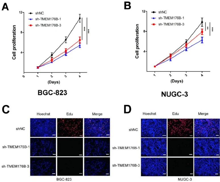
This study investigated the role of the differentially expressed gene TMEM176B in gastric cancer (GC). Western blotting and qPCR analysis were used to assess TMEM176B expression in normal human gastric mucosa cells (GES-1) and human GC cell lines (BGC-823 and NUGC-3). shRNA-mediated TMEM176B knockdown in cancer cells was used for phenotypic analysis, proliferation assays, and apoptosis experiments.
The results indicated that TMEM176B was highly expressed in GC cell lines. TMEM176B knockdown in GC cell lines inhibited cell proliferation (reduced CCK8 and colony formation), increased apoptosis (higher Bax/Bcl2 ratio), and arrested the cell cycle in the G0/G1 phase. This identified TMEM176B's role in GC development and progression, offering molecular targets and a foundation for future treatments.
Ask a Question
Write your own review
Description: Small cell gastrointestinal carcinoma showing creatine kinase-BB and aromatic L-amino-acid decarboxy. Cell growth is slow.

Description: Human cell line derived from signet ring cell carcinoma of stomach.


Description: Human cell line derived from stomach cancer (gastric cancer; highly differentiated tubular adenocarcinoma).

Description: Human gastric cancer cell line derived from metastasis at liver.
- Adipose Tissue-Derived Stem Cells
- Human Neurons
- Mouse Probe
- Whole Chromosome Painting Probes
- Hepatic Cells
- Renal Cells
- In Vitro ADME Kits
- Tissue Microarray
- Tissue Blocks
- Tissue Sections
- FFPE Cell Pellet
- Probe
- Centromere Probes
- Telomere Probes
- Satellite Enumeration Probes
- Subtelomere Specific Probes
- Bacterial Probes
- ISH/FISH Probes
- Exosome Isolation Kit
- Human Adult Stem Cells
- Mouse Stem Cells
- iPSCs
- Mouse Embryonic Stem Cells
- iPSC Differentiation Kits
- Mesenchymal Stem Cells
- Immortalized Human Cells
- Immortalized Murine Cells
- Cell Immortalization Kit
- Adipose Cells
- Cardiac Cells
- Dermal Cells
- Epidermal Cells
- Peripheral Blood Mononuclear Cells
- Umbilical Cord Cells
- Monkey Primary Cells
- Mouse Primary Cells
- Breast Tumor Cells
- Colorectal Tumor Cells
- Esophageal Tumor Cells
- Lung Tumor Cells
- Leukemia/Lymphoma/Myeloma Cells
- Ovarian Tumor Cells
- Pancreatic Tumor Cells
- Mouse Tumor Cells