

Immortalized Human Gingival Keratinocytes-SV40
Cat.No.: CSC-I2301Z
Morphology: Polygonal
Culture Properties: Adherent
- Specification
- Background
- Scientific Data
- Q & A
- Customer Review
Immortalized human gingival keratinocytes-SV40 (HGK/SV40) are generated by introducing the SV40 large T antigen into primary human gingival keratinocytes. This confers indefinite proliferative capacity while preserving key keratinocyte features. In low-calcium serum-free medium, HGK/SV40 maintain a basal/proliferative phenotype expressing p63, cytokeratin 5/14, and E-cadherin. Upon calcium switch or air-liquid interface culture, they can differentiate toward a stratified squamous phenotype with cytokeratin 13, involucrin, and junctional proteins such as ZO-1 and occludin, supporting studies of oral epithelial barrier formation and differentiation.
Compared with primary human gingival keratinocytes, HGK/SV40 offer a continuous, scalable, and cost-effective source with high batch-to-batch consistency and reduced donor variability. This enables reproducible high-throughput screening, long-term culture, and siRNA/CRISPR perturbation. They retain innate immune responsiveness through Toll-like receptors and respond to inflammatory stimuli such as IL-1β, TNF-α, and bacterial LPS, with cytokine and chemokine secretion relevant to periodontal immunity. They are widely used to model periodontal inflammation, peri-implantitis, oral mucositis, wound healing, and host–microbiome interactions, and to test dental materials, oral care products, and pharmaceuticals for biocompatibility and toxicity.
Fiber Architecture Directs Proliferation and Differentiation in Gingival Keratinocytes
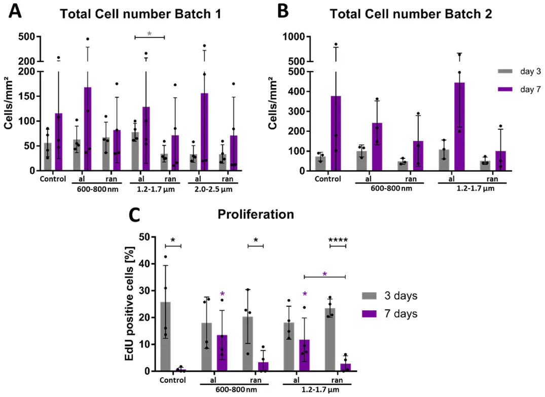
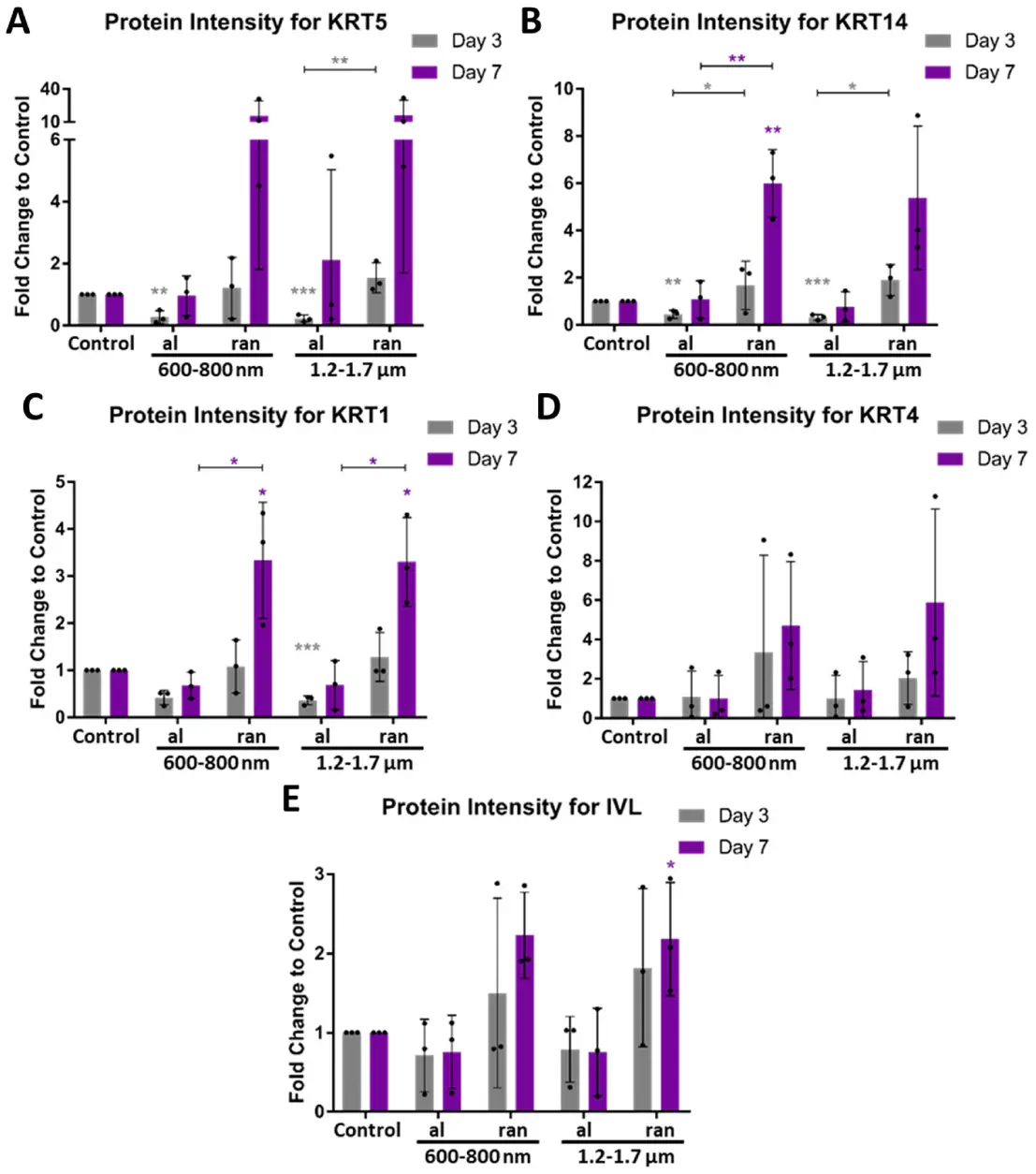
This study systematically evaluated how electrospun polycaprolactone (PCL) scaffolds with defined fiber orientations (aligned vs. random) and diameters (600–800 nm, 1.2–1.7 µm, 2.0–2.5 µm) direct gingival keratinocyte fate. Using immortalized human gingival keratinocytes (ihGK), we assessed cell and nuclear morphology, proliferation dynamics, differentiation marker expression, and the effects of basal keratin (KRT5/KRT14) knockdown.
Quantitative morphological analysis revealed scaffold-dependent changes in cell shape: aligned medium-diameter fibers (with fiber diameters of 1.2–1.7 µm) induced pronounced cell and nuclear elongation, whereas random fibers (600–800 nm) promoted larger, more rounded cell and nuclear shapes. Time-resolved EdU assays indicated that aligned scaffolds supported sustained proliferation, whereas random scaffolds elicited a transient proliferative burst followed by a decline. Gene expression analysis (ddPCR) demonstrated that random scaffolds (especially 600–800 nm fibers) upregulated basal keratins (KRT5, KRT14) and early differentiation markers (KRT1, KRT10, KRT4, KRT13) relative to aligned scaffolds. At the protein level, differentiation markers involucrin (IVL) and filaggrin (FLG) were likewise elevated on random scaffolds, corroborating the mRNA findings. Collectively, these findings establish fiber orientation and diameter as key design parameters for controlling keratinocyte fate.
Ask a Question
Write your own review

Description: The Immortalized Drosophila Embryonic Cells (R1) was established by serial passaging of primary cultures from Act5C > UAS-RasV12embryos. The RasV12immortalized cell lines have significantly ...
Description: Immortalized Cynomolgus Monkey Pulmonary Artery Smooth Muscle Cells-GFP provided by Creative Bioarray have been developed by immortalizing cynomolgus monkey pulmonary artery smooth muscle cells with ...
Description: Immortalized Monkey Spleen Fibroblasts-SV40T were developed from monkey tissues transduced with a lentiviral expression vector containing the SV40T gene. The cell line was continuously cultured for ...
Description: Immortalized Cynomolgus Monkey Prostate Smooth Muscle Cells-GFP provided by Creative Bioarray have been developed by immortalizing cynomolgus monkey prostate smooth muscle cells with SV40 Large T ...
Description: Immortalized Cynomolgus Monkey Spleen Epithelial Cells-GFP provided by Creative Bioarray have been developed by immortalizing cynomolgus monkey spleen epithelial cells with SV40 Large T antigen and ...
Description: Immortalized Mouse Esophageal Epithelial Cells-SV40 have been obtained immortalizing Mouse Esophageal Epithelial Cells with Lenti-SV40 Lentivirus. Immortalized cells were controlled passaging side by ...
- Adipose Tissue-Derived Stem Cells
- Human Neurons
- Mouse Probe
- Whole Chromosome Painting Probes
- Hepatic Cells
- Renal Cells
- In Vitro ADME Kits
- Tissue Microarray
- Tissue Blocks
- Tissue Sections
- FFPE Cell Pellet
- Probe
- Centromere Probes
- Telomere Probes
- Satellite Enumeration Probes
- Subtelomere Specific Probes
- Bacterial Probes
- ISH/FISH Probes
- Exosome Isolation Kit
- Human Adult Stem Cells
- Mouse Stem Cells
- iPSCs
- Mouse Embryonic Stem Cells
- iPSC Differentiation Kits
- Mesenchymal Stem Cells
- Immortalized Human Cells
- Immortalized Murine Cells
- Cell Immortalization Kit
- Adipose Cells
- Cardiac Cells
- Dermal Cells
- Epidermal Cells
- Peripheral Blood Mononuclear Cells
- Umbilical Cord Cells
- Monkey Primary Cells
- Mouse Primary Cells
- Breast Tumor Cells
- Colorectal Tumor Cells
- Esophageal Tumor Cells
- Lung Tumor Cells
- Leukemia/Lymphoma/Myeloma Cells
- Ovarian Tumor Cells
- Pancreatic Tumor Cells
- Mouse Tumor Cells