
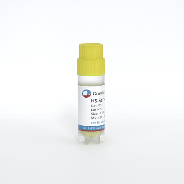
HS-Sch-2
Cat.No.: CSC-C6318J
Species: Homo sapiens (Human)
Morphology: other
Culture Properties: Adherent cells
- Specification
- Background
- Scientific Data
- Q & A
- Customer Review
Store in liquid nitrogen.
HS-Sch-2 cell line was developed from a malignant peripheral nerve sheath tumor (MPNST) in 54 years old woman. It was first reported by McCluggage et al., who morphologically, cytogenetically, and molecularly characterized it as a cell line with stable long-term growth in vitro and complex chromosomal alterations frequently found in aggressive tumors. HS-Sch-2 cells are adherent cells that maintain most of the histopathologic features of the original tumor. The cells express neural crest-derived tumors markers, such as S-100 protein and neuron-specific enolase. HS-Sch-2 has been found to harbor a p53 point mutation (codon 273), mutations commonly found in tumor progression.
More recent genomic studies have led to the suggestion that HS-Sch-2 might actually originate from a desmoplastic melanoma. It is useful for studying both melanoma biology and peripheral nerve sheath tumors. The cell line is reported to have mutations in NF1 as well as deletions in CDKN2A. HS-Sch-2 cells are often used in studies involving tumor biology, oncogenic pathways, and drug discovery.
The Combination of Genome, Methylome, and Expression Marker Analysis, Represent Useful Tools for a Better Differential Diagnosis and Classification of Malignant Peripheral Nerve Sheath Tumors
Malignant peripheral nerve sheath tumors (MPNSTs) are soft-tissue sarcomas of the peripheral nervous system that develop either sporadically or in the context of neurofibromatosis type 1 (NF1). MPNST diagnosis is challenging and treatment outcomes are poor. Magallón-Lorenz et al. present here a resource consisting of the genomic characterization of 9 widely used human MPNST cell lines for translational research.
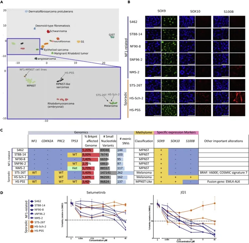
Genomic analysis revealed considerable diversity among cell lines, prompting further characterization. Methylome analysis compared all cell lines to reference cohorts of peripheral nerve sheath and soft tissue tumors to clarify diagnostic classification. UMAP analysis showed all NF1-related MPNST cell lines clustered within the MPNST group, whereas sporadic cell lines fell into the melanoma cluster (STS-26T, HS-Sch-2) or MPNST-like sarcomas (HS-PSS) (Fig. 1A). Immunostaining for SOX9, SOX10, and S100B further characterized 8 cell lines. SOX9 was expressed in all lines. All NF1-related lines and STS-26T were negative for SOX10 and S100B, whereas HS-PSS and HS-Sch-2 were SOX10-positive, and HS-Sch-2 additionally expressed S100B (Fig. 1B)-markers consistent with melanoma rather than classic MPNST identity.
Ask a Question
Write your own review
- You May Also Need
Description: Subline of GOTO. Protein-free medium adapted. Cell growth is slow.
Description: Established from the adrenal tumor tissue resected after treatment from a 20-month-old boy of European origin with neuroblastoma (stage III) in 1991
Description: established from the bone marrow metastasis of a 1-year-old female patient with adrenal gland neuroblastoma negative
Description: The line is resistant to infection and focus formation by ecotropic MuLV.
Description: Established from the primary adrenal tumor resected prior to treatment from a 3-year-old boy with rapidly progressing Stage III neuroblastoma in 1989; described to lack MYCN (NMYC, N-myc) ...
- Adipose Tissue-Derived Stem Cells
- Human Neurons
- Mouse Probe
- Whole Chromosome Painting Probes
- Hepatic Cells
- Renal Cells
- In Vitro ADME Kits
- Tissue Microarray
- Tissue Blocks
- Tissue Sections
- FFPE Cell Pellet
- Probe
- Centromere Probes
- Telomere Probes
- Satellite Enumeration Probes
- Subtelomere Specific Probes
- Bacterial Probes
- ISH/FISH Probes
- Exosome Isolation Kit
- Human Adult Stem Cells
- Mouse Stem Cells
- iPSCs
- Mouse Embryonic Stem Cells
- iPSC Differentiation Kits
- Mesenchymal Stem Cells
- Immortalized Human Cells
- Immortalized Murine Cells
- Cell Immortalization Kit
- Adipose Cells
- Cardiac Cells
- Dermal Cells
- Epidermal Cells
- Peripheral Blood Mononuclear Cells
- Umbilical Cord Cells
- Monkey Primary Cells
- Mouse Primary Cells
- Breast Tumor Cells
- Colorectal Tumor Cells
- Esophageal Tumor Cells
- Lung Tumor Cells
- Leukemia/Lymphoma/Myeloma Cells
- Ovarian Tumor Cells
- Pancreatic Tumor Cells
- Mouse Tumor Cells