TM3
Cat.No.: CSC-C9738L
Species: Mus musculus (Mouse)
Source: Testis
Morphology: epithelial
Culture Properties: monolayer
- Specification
- Background
- Scientific Data
- Q & A
- Customer Review
Strain: BALB/c nu/+
Receptor: luteinizing hormone (LH); epidermal growth factor(EGF); androgen; estrogen; progesterone
Production: prostaglandin F2a
Histopathology: normal
Note: the TM3 cell line responds to LH with an increase in cAMP production, but does not respond to follicle stimulating hormone (FSH); the maintenance of responsiveness to LH is dependent upon serum lot; in the presence of LH, the cells are capable of metabolizing cholesterol; tested and found negative for ectromelia virus (mousepox)
TM3 cells were originally derived from normal testicular tissue obtained from an immature BALB/c mouse. As Leydig cells synthesize the majority of testosterone within the testis, TM3 cells provide an appropriate cell line with which to study steroidogenesis and androgen regulation in vitro.
TM3 cells adhere to culture plates as fibroblast-like cells. They have been shown to express multiple enzymes essential for steroidogenesis including 3β-hydroxysteroid dehydrogenase (3β-HSD). While TM3 cells have relatively low basal levels of testosterone secretion when compared to primary Leydig cells, they have been successfully utilized in mechanistic studies as they can be hormonally stimulated and subjected to signaling pathway alterations. These cells are typically cultured in an equal mixture of DMEM and Ham's F-12 with serum.
Due to their derivation from Leydig cells, TM3 cells have been widely used to study male reproductive physiology, endocrine disruption, and toxicology. Specifically, they have been employed to assess cytotoxicity, steroidogenic potential, and cellular stress responses of environmental chemicals, pharmaceuticals, and hormonal regulators. TM3 cells have proven useful to study Leydig cell biology due to their robustness, ease of culture, and well-established phenotype.
Toxic Effect of BPA Exposure on TM3 Cells
BPA is known to be an endocrine disruptor and has been shown to be reproductive toxicants. However, the mechanism underlying BPA-induced reproductive toxicity in Leydig cells has not been determined. Yang's team pretreated TM3 cells with BPA and then treated with ROS scavenger (N-acetylcysteine), Caspase-3 inhibitor (Ac-DEVD-CHO), autophagy activator (Torin2) or autophagy inhibitor (Chloroquine) to determine the potential mechanism involved.
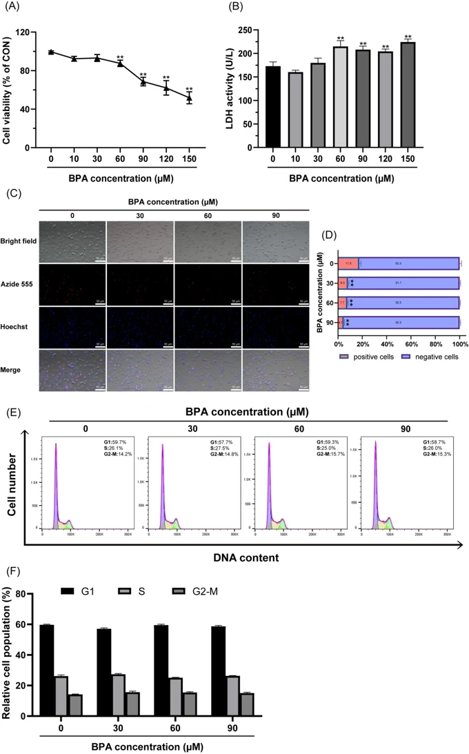
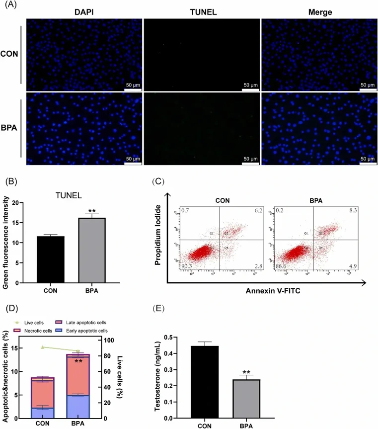
Results from screening studies revealed that treatment of TM3 cells with BPA at ≥60 µM significantly decreased cell viability (Fig. 1A) and proliferation (Fig. 1C and D), but had no significant effect on the cell cycle (Fig. 1E and F). Therefore, they used 60 µM BPA in subsequent experiments. Their study also found that BPA exposure significantly increased apoptosis in TM3 cells using TUNEL staining (Fig. 2A and B) and an increased frequency of annexin V-positive TM3 cells (Fig. 2C and D). ELISA revealed that BPA exposure significantly reduced testosterone synthesis in TM3 cells compared to the control group (Fig. 2E). Taken together, these results indicate that 60 µM BPA is toxic to TM3 cells.
Ask a Question
Write your own review
- You May Also Need

Description: Described as secreting a mouse monoclonal antibody (IgG2a) detecting all fibers in skeletal muscle and myosin heavy chains on Western blots and detecting mammalian, chicken, zebrafish, axolotl, ...

Description: Animals were immunized with the B6.1 mouse cytotoxic T cell line.


Description: neuroglial and neuronal character coexpressing ependymoma cell line.

Description: Established by irradiation of the adherent cells in long-term bone marrow cultures derived from C3H/HeNSlc strain mice
- Adipose Tissue-Derived Stem Cells
- Human Neurons
- Mouse Probe
- Whole Chromosome Painting Probes
- Hepatic Cells
- Renal Cells
- In Vitro ADME Kits
- Tissue Microarray
- Tissue Blocks
- Tissue Sections
- FFPE Cell Pellet
- Probe
- Centromere Probes
- Telomere Probes
- Satellite Enumeration Probes
- Subtelomere Specific Probes
- Bacterial Probes
- ISH/FISH Probes
- Exosome Isolation Kit
- Human Adult Stem Cells
- Mouse Stem Cells
- iPSCs
- Mouse Embryonic Stem Cells
- iPSC Differentiation Kits
- Mesenchymal Stem Cells
- Immortalized Human Cells
- Immortalized Murine Cells
- Cell Immortalization Kit
- Adipose Cells
- Cardiac Cells
- Dermal Cells
- Epidermal Cells
- Peripheral Blood Mononuclear Cells
- Umbilical Cord Cells
- Monkey Primary Cells
- Mouse Primary Cells
- Breast Tumor Cells
- Colorectal Tumor Cells
- Esophageal Tumor Cells
- Lung Tumor Cells
- Leukemia/Lymphoma/Myeloma Cells
- Ovarian Tumor Cells
- Pancreatic Tumor Cells
- Mouse Tumor Cells