
COLO 201
Cat.No.: CSC-C9361L
Species: Homo sapiens (Human)
Source: Ascites Metastasis
Morphology: fibroblast like ; bipolar, slightly refractile
Culture Properties: cells grow loosely attached and in suspension
- Specification
- Background
- Scientific Data
- Q & A
- Customer Review
Tumorigenecity: yes, in nude mice
Isoenzyme: G6PD,B;PGM1,1-2;PGM3,1-2;6PGD,A;ES-D,1-2;PEP-D,1
Histopathology: adenocarcinoma
Note: CSAp negative (CSAp-); CEA, negative; the line was derived from tissue from the same patient as COLO 205 (KCLB 222)
vWA: 15
FGA: 21,23
Amelogenin: X
TH01: 8,9
TPOX: 11
CSF1P0: 11,12
D5S818: 10,13
D13S317: 10,12
D7S820: 9,10
COLO 201 is a human colorectal adenocarcinoma cell line originally derived from the ascitic fluid of a 70-year-old Caucasian male with Dukes' type D colon cancer. It is a sibling cell line of COLO 205 but shows a different, fibroblast-like, bipolar and slightly refractile appearance. In contrast to many other epithelial colon cancer lines, COLO 201 cells grow almost exclusively in suspension, with only a small proportion of loosely adherent cells. This characteristic may be because COLO 201 cells were isolated from a metastatic ascites sample and not from a solid tumor. COLO 201 is hypertriploid cytogenetically with most cells having 10-11 marker chromosomes (monosomic, disomic or trisomic) and a doubling time of about 41.3 hours. COLO 201 cells are MSS.
COLO 201 expresses the oncogenes MYC, RAS, MYB, FOS, SIS, and mutant p53 and is negative for classical colon tumor markers CEA (carcinoembryonic antigen) and CSAp. The isoenzyme profile (G6PD, PGM, PGD) of the cell line has been fully characterized, and has been used to authenticate species and cell-line purity. COLO 201 is highly tumorigenic: intraperitoneal injection of 107 cells into immunodeficient (nude) mice leads to 100% engraftment and tumor formation after 21 days. This cell line is used as an experimental model in many cancer biology, toxicology, high-throughput screening, and three-dimensional cell culture studies.
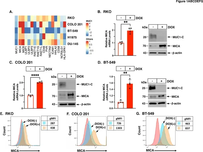
MUC1-C Suppresses MICA Expression by Human Cancer Cells
MUC1-C evolved to protect barrier tissues but promotes oncogenesis and T cell dysfunction in cancers. Its role in NK cell function is unknown. Morimoto et al. investigated how MUC1-C affects NK cell-activating MICA/B ligands and explores mechanisms of immune evasion in cancer.
To test whether MUC1-C has a role in innate NK cell immunity, we assessed its impact on the expression of NKG2D ligands. RNA-seq data revealed that silencing MUC1-C significantly upregulated MICA and MICB transcripts in human colorectal cancer (RKO, COLO 201), triple-negative breast cancer (BT-549), non-small cell lung cancer (H1975), and castration-resistant prostate cancer (DU-145) cells (Fig. 1A). Silencing MUC1-C also increased ULBP3, ULBP6/RAET1L, and CD48 expression, though the effects on ULBP ligands varied across cancer types (Fig. 1A). They focused on MICA and confirmed that silencing MUC1-C induced MICA expression in RKO (Fig. 1B) and COLO 201 cells (Fig. 1C). Similar results were seen with a second MUC1 shRNA. MUC1-C was also required for MICA expression in BT-549 (Fig. 1D), H1975, and DU-145 cells (online supplemental figure S1C). Flow cytometry analyses showed that MUC1 silencing led to increased cell surface MICA in RKO (Fig. 1E) and COLO 201 cells (Fig. 1F) and yielded similar results in BT-549 (Fig. 1G), H1975, and DU-145 cells. Taken together, MUC1-C represses MICA gene and cell surface expression in a wide range of cancer cells.
Ask a Question
Write your own review
- You May Also Need

Description: Established from an adenocarcinoma located in the ascending colon of a 68 year-old male patient. The adenocarcinoma was well differentiated and determined to be Dukes' stage B. Imperial College ...

Description: This is one cell line out of a series of colon carcinoma cell lines established by PD Dr. Michael Linnebacher.

Description: Rectal carcinoma from a Japanese patient. Cell growth is slow.

Description: The C80 cell line was established from a 69-year old male patient with a moderately well differentiated adenocarcinoma of the rectum classified as Dukes' stage D.


Description: RKO is a poorly differentiated colon carcinoma cell line developed by Michael Brattain.
- Adipose Tissue-Derived Stem Cells
- Human Neurons
- Mouse Probe
- Whole Chromosome Painting Probes
- Hepatic Cells
- Renal Cells
- In Vitro ADME Kits
- Tissue Microarray
- Tissue Blocks
- Tissue Sections
- FFPE Cell Pellet
- Probe
- Centromere Probes
- Telomere Probes
- Satellite Enumeration Probes
- Subtelomere Specific Probes
- Bacterial Probes
- ISH/FISH Probes
- Exosome Isolation Kit
- Human Adult Stem Cells
- Mouse Stem Cells
- iPSCs
- Mouse Embryonic Stem Cells
- iPSC Differentiation Kits
- Mesenchymal Stem Cells
- Immortalized Human Cells
- Immortalized Murine Cells
- Cell Immortalization Kit
- Adipose Cells
- Cardiac Cells
- Dermal Cells
- Epidermal Cells
- Peripheral Blood Mononuclear Cells
- Umbilical Cord Cells
- Monkey Primary Cells
- Mouse Primary Cells
- Breast Tumor Cells
- Colorectal Tumor Cells
- Esophageal Tumor Cells
- Lung Tumor Cells
- Leukemia/Lymphoma/Myeloma Cells
- Ovarian Tumor Cells
- Pancreatic Tumor Cells
- Mouse Tumor Cells