
CAL-39
Cat.No.: CSC-C0316
Species: Homo sapiens (Human)
Source: Vulva
Morphology: adherent, epithelial-like cells growing as monolayer
Culture Properties: monolayer
- Specification
- Background
- Scientific Data
- Q & A
- Customer Review
Immunology: cytokeratin +, desmin -, endothel -, GFAP -, neurofilament -, vimentin -
Viruses: ELISA: reverse transcriptase negative; PCR: EBV -, HBV -, HCV -, HHV-8 -, HIV -,
CAL-39 was derived from primary bladder tumor and is a human urothelial carcinoma cell line. As such, CAL-39 cells are used as an in vitro model system for understanding bladder cancer biology. These cells maintain many molecular and phenotypic features of urothelial carcinoma. They are able to form adherent epithelial monolayers in culture and have a polygonal shape that is characteristic of urothelial tumor cells. They express epithelial markers such as cytokeratins and show altered regulation of cell cycle- and survival-related pathways commonly associated with bladder cancer progression. CAL-39 has been reported to harbor genetic alterations affecting tumor suppressor and oncogenic signaling, and it has been included in multiple pharmacological and molecular studies investigating DNA damage responses, apoptosis, and therapeutic sensitivity. Previous studies have utilized CAL-39 to evaluate the effects of chemotherapeutic agents and targeted compounds, supporting its value in drug screening and resistance research.
CAL-39 cells have been used to study regulation of growth in urothelial carcinoma, deregulation of signaling pathways in bladder cancer, and stress responses in tumor cells. Researchers have also used CAL-39 for comparative analysis with other bladder cancer cell lines.
G1 Acts through GPER1 in Vulvar Carcinoma Cells A431/CAL-39
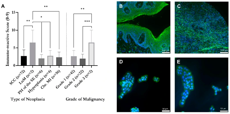
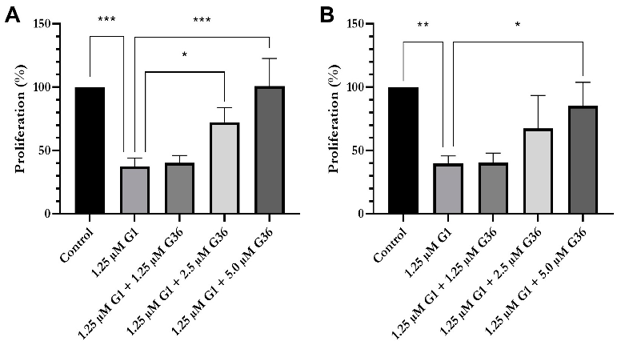
GPER1 can act as either tumor-promoting or tumor-suppressive depending on cancer type, yet its role in vulvar carcinoma remains undefined. Loris et al. clarified whether GPER1 supports or suppresses vulvar cancer progression. First, they determined subcellular localization of GPER1 in vulvar carcinoma cell lines A431/CAL-39, scored expression in a vulvar-neoplasia tissue microarray (TMA), and finally evaluated proliferation, viability, migration, clonogenicity, and morphology after treatment with GPER1 agonist (G1) or antagonist (G36).
Immunofluorescence confirmed GPER1 localization to both the nucleus and cytoplasm with no statistical difference between A431 and CAL-39 vulvar carcinoma cells (Fig. 1D, E). Treatment cells were treated with G1 (1.25 µM) ± varying doses of GPER1 antagonist G36. G1 treatment alone significantly decreased proliferation of both A431 (37.39 ± 3.88% vs. control; p < 0.001; Fig. 2A) and CAL-39 (39.91 ± 3.39% vs. control; p < 0.01; Fig. 2B), and this suppression was reversed in a dose-dependent manner by G36. In A431 cells, proliferation rates rose to 72.34 ± 6.62% (p = 0.05) and 100.65 ± 12.64% (p = 0.001) treated with 2.5 µM and 5 µM G36, respectively. Similarly, CAL-39 proliferation rates showed a dose-dependent recovery to 67.44 ± 15.04% and 85.18 ± 10.71% (p < 0.05) treated with 2.5 and 5 µM G36, respectively.
Ask a Question
Write your own review
- You May Also Need
Description: Established in 2007 from the large retrosternal mass resected before treatment from a 57-year-old Caucasian man with rapidly fatal anaplastic thyroid cancer

Description: established from the uvea tissue of a male patient with uveal melanoma

Description: NTERA-2 was cloned from cell line TERA-2 which was derived from a metastatic teratocarcinoma of a 22-year-old Caucasian male; cell line also known as NT-2
Description: Established from the bone marrow of a 27-year-old woman with B-cell non-Hodgkin lymphoma (B-NHL) (diffuse large cell lymphoma, DLCL, stage 4B, at relapse) in 1987
- Adipose Tissue-Derived Stem Cells
- Human Neurons
- Mouse Probe
- Whole Chromosome Painting Probes
- Hepatic Cells
- Renal Cells
- In Vitro ADME Kits
- Tissue Microarray
- Tissue Blocks
- Tissue Sections
- FFPE Cell Pellet
- Probe
- Centromere Probes
- Telomere Probes
- Satellite Enumeration Probes
- Subtelomere Specific Probes
- Bacterial Probes
- ISH/FISH Probes
- Exosome Isolation Kit
- Human Adult Stem Cells
- Mouse Stem Cells
- iPSCs
- Mouse Embryonic Stem Cells
- iPSC Differentiation Kits
- Mesenchymal Stem Cells
- Immortalized Human Cells
- Immortalized Murine Cells
- Cell Immortalization Kit
- Adipose Cells
- Cardiac Cells
- Dermal Cells
- Epidermal Cells
- Peripheral Blood Mononuclear Cells
- Umbilical Cord Cells
- Monkey Primary Cells
- Mouse Primary Cells
- Breast Tumor Cells
- Colorectal Tumor Cells
- Esophageal Tumor Cells
- Lung Tumor Cells
- Leukemia/Lymphoma/Myeloma Cells
- Ovarian Tumor Cells
- Pancreatic Tumor Cells
- Mouse Tumor Cells