BRF41
Cat.No.: CSC-C9063H
Species: Danio rerio (Zebrafish)
Source: Fin
Morphology: Fibroblast-like
- Specification
- Background
- Scientific Data
- Q & A
- Customer Review
BRF41 (abbreviated from Cell Line of fish Brain cell line 41) is an immortalized cell line derived from the teleost fish Danio rerio (zebrafish). The cell line was isolated from fin tissue and since its isolation has been cultured continuously. As such, BRF41 is a spontaneously immortalized fish cell line. This cell line has been used as a peripheral cell model and has been used for studies in various fields of cellular and molecular biology.
Cells from this line can be grown at intermediate temperatures (~27 °C) in Leibovitz's L‑15 with fetal bovine serum media, where they display fibroblast-like qualities and are capable of continuous growth. BRF41 cells have been used as a cellular model to study various cellular processes. Gene expression analysis revealed that the transcription factors zDEC1 and zDEC2 oscillate during circadian rhythms in BRF41 cells, suggesting their involvement with established feedback loops controlling circadian rhythms. Polyamine metabolism has also been studied in BRF41 cells, determining enzyme kinetics of ornithine decarboxylase in zebrafish. Stress within the endoplasmic reticulum (ER) has also been studied using BRF41 cells. Treatment with tributyltin induces ER stress in zebrafish, which can be analyzed using BRF41 cells.
Release and Degradation of Dissolved Environmental RNAs from zebrafish Cells
The sources and degradation profiles of dissolved environmental RNAs from fish in water are unknown. Xu's team investigated the permeability of RNA from zebrafish cells, the release of dissolved RNAs from live and dying cells, and RNA degradation in a non-sterile aqueous environment to provide insights into fish RNAs in water.
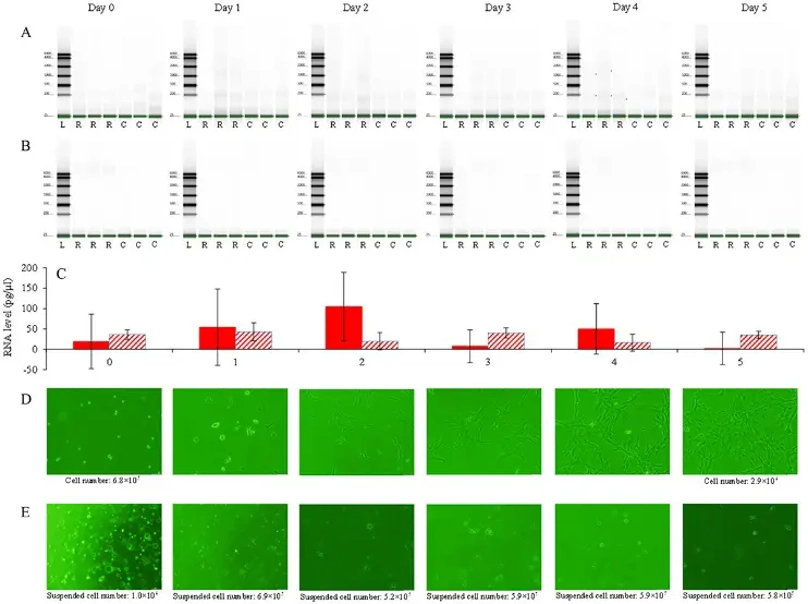
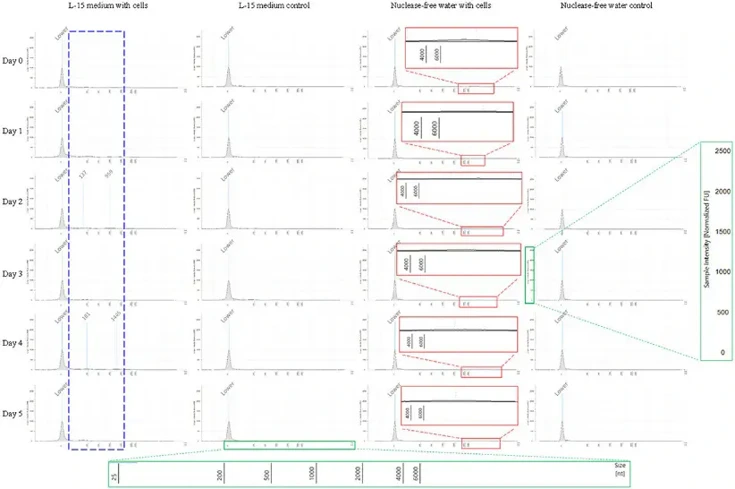
Dissolved RNAs from live zebrafish BRF41 cells in L-15 medium and dying cells in nuclease-free water were detected (Fig. 1A-C). Over 6 days, levels of dissolved RNAs from both types of cells fluctuated (Fig. 1C). RNAs from live cells were mostly under 2000 bases, while those from dying cells were mostly over 4000 bases (Fig. 2). Cells in L-15 medium grew well, mostly attaching to the flask bottom. On day 1, they stretched, and their number tripled by the end of the experiment (Fig. 1D). In contrast, cells in nuclease-free water were dying, with few attaching to the bottom, no growth, and abnormal morphology starting on day 3 (Fig. 1E).
Ask a Question
Write your own review
- You May Also Need


Description: Derived from tissue posterior to the anus of normal adult minnows. Reported to be capable of growth over a wide temperature range from 4°C-36°C with maximum growth at 34°C. Support growth of the ...

Description: E11 is a clone of the cell line SSN-1 and is persistantly infected with a C-type retrovirus (SnRV). It is susceptible to piscine nodavirus strains belonging to different genotypes (SJNNV, RGNNV, ...

Description: The fish cell line SSN-1 was initiated from whole fry tissue, Channa (Ophicephalus) striatus, commonly named 'striped snakehead'. SSN-1 spontaneously produce and release endogenous snakehead fish ...

Description: From pooled male/female gonad tissue of yearling rainbow trout (Oncorhynchus mykiss). First cell line to be established from cold-blooded vertebrates. 8-fold difference in growth rate in temperature ...

- Adipose Tissue-Derived Stem Cells
- Human Neurons
- Mouse Probe
- Whole Chromosome Painting Probes
- Hepatic Cells
- Renal Cells
- In Vitro ADME Kits
- Tissue Microarray
- Tissue Blocks
- Tissue Sections
- FFPE Cell Pellet
- Probe
- Centromere Probes
- Telomere Probes
- Satellite Enumeration Probes
- Subtelomere Specific Probes
- Bacterial Probes
- ISH/FISH Probes
- Exosome Isolation Kit
- Human Adult Stem Cells
- Mouse Stem Cells
- iPSCs
- Mouse Embryonic Stem Cells
- iPSC Differentiation Kits
- Mesenchymal Stem Cells
- Immortalized Human Cells
- Immortalized Murine Cells
- Cell Immortalization Kit
- Adipose Cells
- Cardiac Cells
- Dermal Cells
- Epidermal Cells
- Peripheral Blood Mononuclear Cells
- Umbilical Cord Cells
- Monkey Primary Cells
- Mouse Primary Cells
- Breast Tumor Cells
- Colorectal Tumor Cells
- Esophageal Tumor Cells
- Lung Tumor Cells
- Leukemia/Lymphoma/Myeloma Cells
- Ovarian Tumor Cells
- Pancreatic Tumor Cells
- Mouse Tumor Cells