
- Specification
- Background
- Scientific Data
- Q & A
- Customer Review
vWA: 16,17
FGA: 20,23
Amelogenin: X
TH01: 8
TPOX: 8,10
CSF1P0: 11,12
D5S818: 12
D13S317: 11
D7S820: 9
Shipping Condition: Room Temperature
A375SM is a highly metastatic subline derived from the human malignant melanoma cell line A375. It was established through in vivo selection in nude mice, specifically by selecting for cells with a higher propensity for lung metastasis. Cells in A375SM line show increased migratory and invasive behavior, upregulation of epithelial-mesenchymal transition (EMT) markers such as N-cadherin and Vimentin, and downregulation of E-cadherin. They also secrete vascular endothelial growth factor (VEGF), promoting angiogenesis in the tumor microenvironment. Additionally, A375SM cells often display partial resistance to certain chemotherapeutic drugs like dacarbazine. On a molecular level, they commonly exhibit the BRAF V600E mutation, leading to the activation of the MAPK/ERK signaling pathway, and have active PI3K/AKT signaling. Researchers often utilize this cell line for studying tumor biology and metastasis along with testing drug candidates and evaluating immunotherapy effectiveness. A375SM cells serve as a valuable model to investigate how tumor cells penetrate the basement membrane to spread through the bloodstream and establish distant metastases.
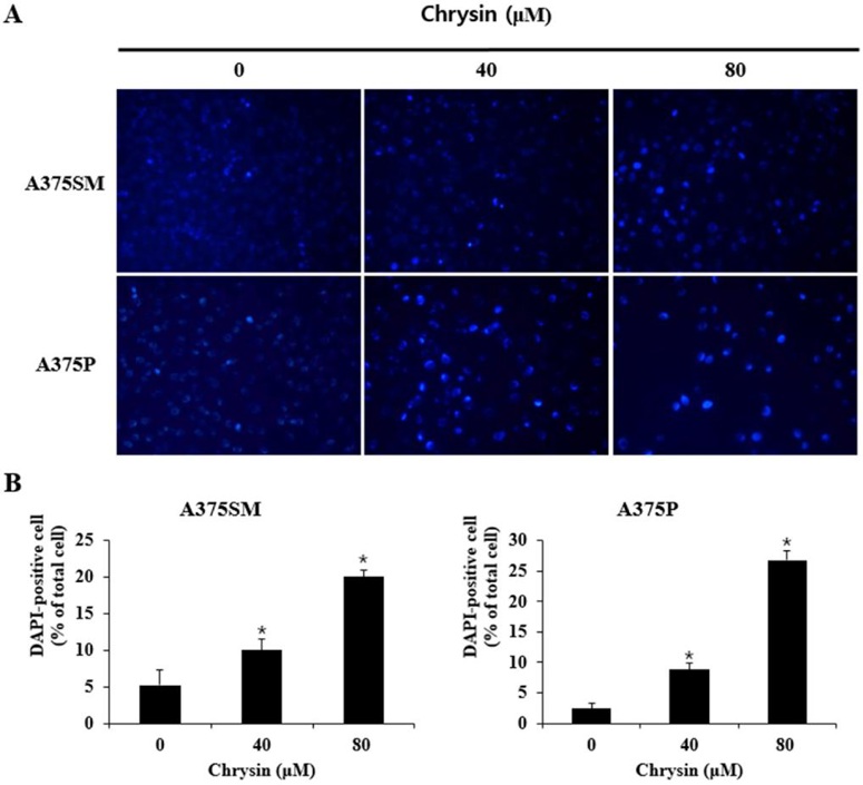
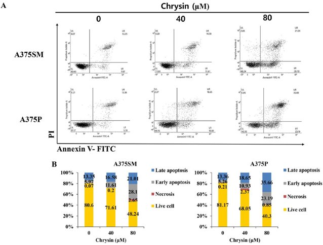
Chrysin-Induced Apoptosis of A375SM and A375P Melanoma Cells
Chrysin is known to exert anti-inflammatory, antioxidant, and anticancer effects. Lee et al. investigated the anticancer effects of chrysin in the human melanoma cells A375SM and A375P.
MTT results confirmed that chrysin decreased the A375SM and A375P melanoma cells' viability. DAPI staining was performed to determine if chrysin-induced decrease in A375SM and A375P cell viability is apoptosis-dependent by examining nuclear morphology. Figure 2A shows nuclear and chromatin condensation in chrysin-treated groups (especially at 100 pM), unlike the control. The number of DAPI-positive cells increased to 5.24%, 10.02%, and 20.02% in A375SM and 2.37%, 8.86%, and 26.83% in A375P with 0, 40, and 80 μM, respectively (Fig. 1B). Flow cytometry by annexin V/PI staining confirmed that apoptosis (early/late) increased in a concentration-dependent manner (Fig. 2A). The total apoptosis was observed to increase to 19.32%, 28.19%, and 39.71% in A375SM and 18.62%, 29.58%, and 58.85% in A375P (Fig. 2B). Additionally, western blot was performed to confirm the increased expression of Bax (proapoptotic), decreased expression of Bcl-2 (anti-apoptotic) and PARP/caspase substrate fragmentation, confirming apoptosis-mediated proliferation inhibition.
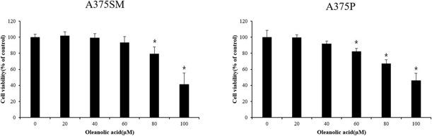
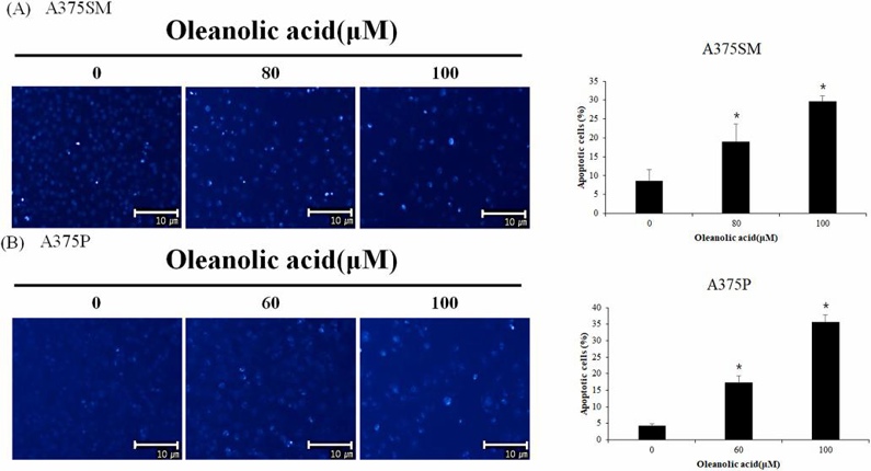
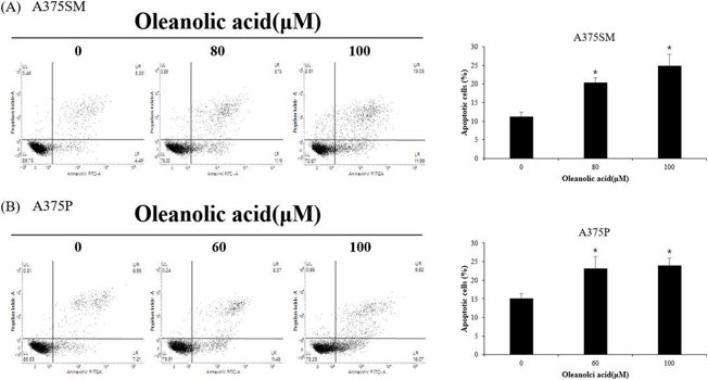
Oleanolic Acid Reduced Cell Viability and Induced Apoptosis of A375SM and A375P Cells
Owing to the ineffectiveness of the currently used therapies against melanoma, there has been a shift in focus toward alternative therapies involving the use of natural compounds. Woo et al. assessed the anticancer effects of oleanolic acid (OA) and its ability to induce apoptosis in A375SM and A375P melanoma cells in vivo.
The MTT assay revealed that OA decreased the viability of A375SM and A375P melanoma cells in a concentration-dependent manner. In A375SM, the viability was 79.6% and 41.5% at 80 μM and 100 μM OA, respectively, and in A375P, it was 82.4% and 46.4% at 60 μM and 100 μM OA, respectively (Fig. 3). DAPI staining indicated an increase in DAPI-positive cells in A375SM (8.7%, 19.0%, 29.7%) and A375P (4.3%, 17.3%, 35.7%) at 0, 80/60, and 100 μM OA, respectively (Fig. 4). Flow cytometry analysis confirmed the apoptosis induction, and the apoptotic rates were 11.1%, 20.4%, and 24.9% in A375SM (0, 80, 100 μM OA) and 15.0%, 23.2%, and 23.9% in A375P (0, 60, 100 μM OA) (Fig. 5). These data suggest that OA induces apoptosis in both cell lines.
Ask a Question
Write your own review
- You May Also Need

Description: Established from the primary tumor (right cervical) of a 64-year-old woman with cutaneous melanoma

Description: Established from the lymph node metastasis of a malignant melanoma from a 42-year-old Caucasian woman
Description: Species: human - male, 31 years old, CaucasianIsoenzyme: G6PD, BProduction: melaninHistopathology: melanoma
Description: Epstein-Barr virus-positive cell line established from peripheral blood in 1977 from male patient with melanoma

Description: Established from the primary tumor of a 58-year-old woman with melanoma in 1977

Description: Established from the primary (achromic) cutaneous tumor (left thigh) of a 26-year-old man with malignant melanoma (primary tumor histology: SSM level IV); same patient as cell line IGR-37; described ...
- Adipose Tissue-Derived Stem Cells
- Human Neurons
- Mouse Probe
- Whole Chromosome Painting Probes
- Hepatic Cells
- Renal Cells
- In Vitro ADME Kits
- Tissue Microarray
- Tissue Blocks
- Tissue Sections
- FFPE Cell Pellet
- Probe
- Centromere Probes
- Telomere Probes
- Satellite Enumeration Probes
- Subtelomere Specific Probes
- Bacterial Probes
- ISH/FISH Probes
- Exosome Isolation Kit
- Human Adult Stem Cells
- Mouse Stem Cells
- iPSCs
- Mouse Embryonic Stem Cells
- iPSC Differentiation Kits
- Mesenchymal Stem Cells
- Immortalized Human Cells
- Immortalized Murine Cells
- Cell Immortalization Kit
- Adipose Cells
- Cardiac Cells
- Dermal Cells
- Epidermal Cells
- Peripheral Blood Mononuclear Cells
- Umbilical Cord Cells
- Monkey Primary Cells
- Mouse Primary Cells
- Breast Tumor Cells
- Colorectal Tumor Cells
- Esophageal Tumor Cells
- Lung Tumor Cells
- Leukemia/Lymphoma/Myeloma Cells
- Ovarian Tumor Cells
- Pancreatic Tumor Cells
- Mouse Tumor Cells