
Strain C57BL/6 Mouse Mesenchymal Stem Cells with GFP
- Specification
- Scientific Data
- Q & A
- Customer Review
Cell patterning in vivo using microrobot specifically designed for tissue engineering applications
Background
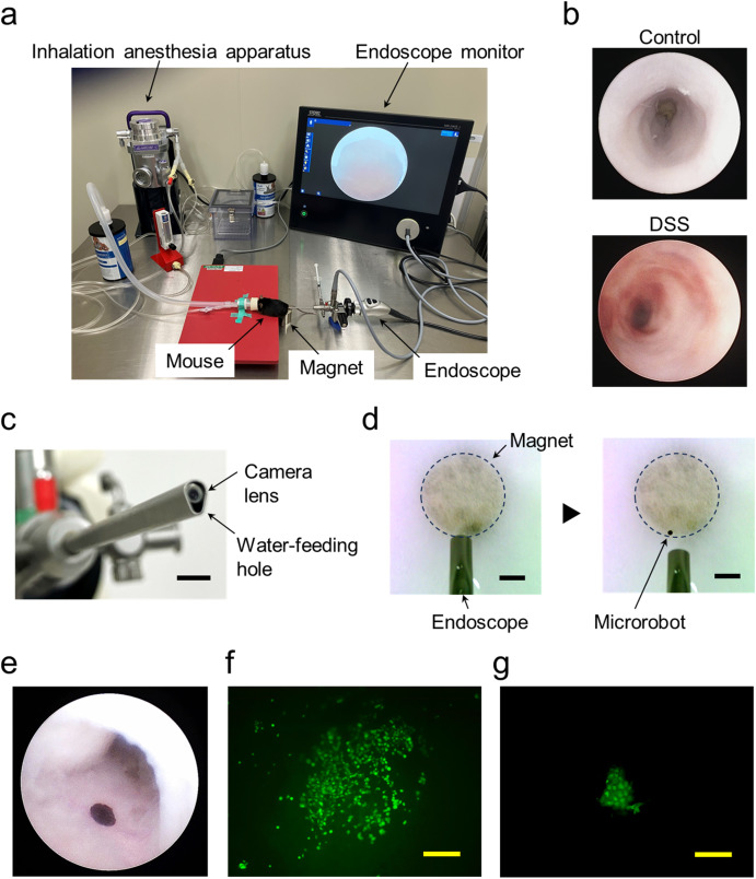
This study introduces a pioneering microrobot-based approach for in vivo cell patterning in tissue engineering. The microrobot, fabricated from serum albumin and magnetic nanoparticles with cell membrane-anchoring reagents, captures cells and rapidly releases them at target sites. Cellular patterns were successfully created on Matrigel, other cell types, and inflamed colon tissues within 30 minutes. As a proof-of-concept, stem cell patterning was performed inside the colons of colitis mice using an endoscope and magnetic guidance, demonstrating persistent cell survival for 9 days. This opens a new field of in vivo cell patterning for advanced stem cell therapies.
Key Findings
The microrobot was fabricated from recombinant human serum albumin, magnetic nanoparticles (MNPs), and cell membrane-anchoring reagents (CMAR), enabling cell capture and rapid release. Optimal CMAR concentrations (0.5 μM for MSCs, 1 μM for NIH3T3 cells) allowed complete cell release within 30 min. Cellular patterns of NIH3T3 cells and MSCs were successfully created on Matrigel (83% and 67% coverage, respectively) and on other cell types. On inflamed colon tissues, MSC patterns were formed ex vivo. In vivo, MSC-loaded microrobots were delivered via endoscope into colitis mouse colons, guided by an external magnet, and transferred to damaged tissues, creating circular stem cell patterns within 30 min. Patterned MSCs persisted for at least 9 days, demonstrating sustained survival. The microrobot did not degrade rapidly and was excreted, with optional retrieval via magnetic forceps. This first demonstration of in vivo cell patterning shows potential for precise stem cell therapies.
Contribution
Creative Bioarray provided GFP-expressing C57BL/6N mouse bone marrow mesenchymal stem cells (Catalog no. CSC-C1314) and SuperCult® mouse MSC growth medium. These reagents enabled the capture, patterning, and in vivo tracking of MSCs, serving as critical cellular tools for demonstrating microrobot-based cell patterning in colitis mice.
Ask a Question
Write your own review
- Adipose Tissue-Derived Stem Cells
- Human Neurons
- Mouse Probe
- Whole Chromosome Painting Probes
- Hepatic Cells
- Renal Cells
- In Vitro ADME Kits
- Tissue Microarray
- Tissue Blocks
- Tissue Sections
- FFPE Cell Pellet
- Probe
- Centromere Probes
- Telomere Probes
- Satellite Enumeration Probes
- Subtelomere Specific Probes
- Bacterial Probes
- ISH/FISH Probes
- Exosome Isolation Kit
- Human Adult Stem Cells
- Mouse Stem Cells
- iPSCs
- Mouse Embryonic Stem Cells
- iPSC Differentiation Kits
- Mesenchymal Stem Cells
- Immortalized Human Cells
- Immortalized Murine Cells
- Cell Immortalization Kit
- Adipose Cells
- Cardiac Cells
- Dermal Cells
- Epidermal Cells
- Peripheral Blood Mononuclear Cells
- Umbilical Cord Cells
- Monkey Primary Cells
- Mouse Primary Cells
- Breast Tumor Cells
- Colorectal Tumor Cells
- Esophageal Tumor Cells
- Lung Tumor Cells
- Leukemia/Lymphoma/Myeloma Cells
- Ovarian Tumor Cells
- Pancreatic Tumor Cells
- Mouse Tumor Cells