
Sprague-Dawley (SD) Rat Mesenchymal Stem Cells with RFP
- Specification
- Scientific Data
- Q & A
- Customer Review
Abstract No. 314 Pre-operative Portal Vein Ligation and MSC Injection in a Rat Model
Background
Preoperative portal vein ligation (PVL) redirects portal flow to the future liver remnant (FLR) to induce hypertrophy before hepatic resection. This study investigated whether mesenchymal stem cells (MSCs) can augment PVL in a rat model. RFP-labeled Sprague-Dawley rat bone-marrow-derived MSCs were injected into non-occluded portal branches after PVL. CT volumetry, necropsy, and histology assessed FLR growth and MSC presence. MSC addition improved liver hypertrophy, suggesting a potential adjunct strategy for liver cancer resection.
Key Findings
All rats survived for two weeks with no complications. CT volumetry showed that PVL+MSC rats had a 70.2% increase in non-ligated liver volume (1.54 to 3.08 mL), compared with a 41.4% increase in PVL-only rats (1.19 to 1.91 mL) and a decrease in controls (2.99 to 1.35 mL). Necropsy revealed the highest future liver remnant (FLR)-to-total weight ratio in the PVL+MSC group. Histological evaluation confirmed RFP-labeled MSCs only in the PVL+MSC group, not in PVL or control groups. These results indicate that MSC injection can enhance PVL-induced liver hypertrophy in this rat model, supporting further research into MSC retention and clinical translation for liver resection.
Contribution
Creative Bioarray provided the RFP-labeled Sprague-Dawley rat bone-marrow-derived mesenchymal stem cells (MSCs) used in the PVL+MSC group. This material enabled histologic tracking and assessment of MSC-enhanced liver hypertrophy after preoperative portal vein ligation in the rat model, supporting the study's key findings.
Injectable, electrosprayed RGD-coupled alginate hydrogel microcapsules enable enhanced viability and sustained release of mesenchymal stem cells
Background
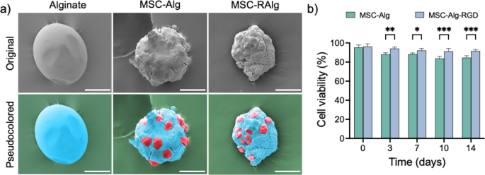
This study develops an injectable cell-delivery platform using electrosprayed RGD-coupled alginate (RAlg) microcapsules to enhance mesenchymal stem cell (MSC) viability and sustained release. Electrospray parameters were optimized to produce uniform microcapsules (~175 µm) with a microporous architecture. RGD conjugation did not significantly alter swelling, viscoelasticity, or encapsulation efficiency. Encapsulated MSCs remained viable, with RAlg maintaining 91.3% viability after 14 days versus 84.9% for alginate alone. Both formulations showed comparable time-dependent cell release, supporting RAlg microcapsules as a biocompatible, injectable platform for regenerative medicine.
Key Findings
Electrospraying parameters were optimized (7 kV, 0.1 mL/min, 80 µm needle, 2% alginate) to fabricate uniform microcapsules with a mean diameter of 175.4 ± 21.1 µm. Electron microscopy revealed a rough surface and an ordered microporous architecture with pores <200 nm. RGD conjugation did not significantly alter swelling, viscoelasticity at low concentrations, or encapsulation efficiency; both alginate and RAlg achieved >92% encapsulation. MSCs were successfully encapsulated and exhibited a round morphology. Over 14 days, RAlg microcapsules maintained significantly higher MSC viability (91.3%) than unmodified alginate (84.9%), with differences emerging by day 3. Both microcapsule types showed comparable time-dependent release: mostly intact at day 1, 59–61% cell release by day 7, and 78–81% release by day 14. These results indicate that electrosprayed RAlg microcapsules provide enhanced viability and sustained release, offering a biocompatible injectable delivery platform for MSC therapies.
Contribution
Creative Bioarray provided the bone marrow-derived Sprague-Dawley red fluorescent protein (RFP)-mesenchymal stem cells (MSCs) used throughout the study. These cells enabled visualization of encapsulation, viability assessment, and release experiments, supporting the evaluation of electrosprayed RAlg microcapsules as an injectable MSC delivery platform.
Ask a Question
Write your own review
- Adipose Tissue-Derived Stem Cells
- Human Neurons
- Mouse Probe
- Whole Chromosome Painting Probes
- Hepatic Cells
- Renal Cells
- In Vitro ADME Kits
- Tissue Microarray
- Tissue Blocks
- Tissue Sections
- FFPE Cell Pellet
- Probe
- Centromere Probes
- Telomere Probes
- Satellite Enumeration Probes
- Subtelomere Specific Probes
- Bacterial Probes
- ISH/FISH Probes
- Exosome Isolation Kit
- Human Adult Stem Cells
- Mouse Stem Cells
- iPSCs
- Mouse Embryonic Stem Cells
- iPSC Differentiation Kits
- Mesenchymal Stem Cells
- Immortalized Human Cells
- Immortalized Murine Cells
- Cell Immortalization Kit
- Adipose Cells
- Cardiac Cells
- Dermal Cells
- Epidermal Cells
- Peripheral Blood Mononuclear Cells
- Umbilical Cord Cells
- Monkey Primary Cells
- Mouse Primary Cells
- Breast Tumor Cells
- Colorectal Tumor Cells
- Esophageal Tumor Cells
- Lung Tumor Cells
- Leukemia/Lymphoma/Myeloma Cells
- Ovarian Tumor Cells
- Pancreatic Tumor Cells
- Mouse Tumor Cells