SKN
Cat.No.: CSC-C6491J
Species: Homo sapiens (Human)
Source: Uterus
Morphology: fibroblast-like
Culture Properties: Adherent cells
- Specification
- Background
- Scientific Data
- Q & A
- Customer Review
Store in liquid nitrogen.
SKN Cells are derived from human tumor uterine corpus leiomyosarcoma. Leiomyosarcoma is a type of cancer that starts in smooth muscles of the uterus. SKN cells provide a platform to study the molecular basis of leiomyosarcoma initiation, progression and treatment response.
In most culture conditions, SKN cells will grow adherently as spindle-shaped cells like other smooth muscle-derived cells. SKN cells express α-smooth muscle actin (α-SMA), desmin and vimentin, markers of smooth muscle cells, demonstrating their mesenchymal nature. SKN cells may also phenotypically possess traits typical of cancer cells with high proliferation ability, cellular atypia, and high invasion capacity.
SKN cells can be used to study tumor cell proliferation, migration, invasion and extracellular matrix remodeling. They are commonly used to study sarcoma-associated signaling such as PI3K/AKT, MAPK, TGF-β, as well as apoptotic resistance and tumor progression. SKN cells are used as models to test chemotherapeutic drugs, targeted therapies and novel anti-cancer drugs against uterine leiomyosarcoma.
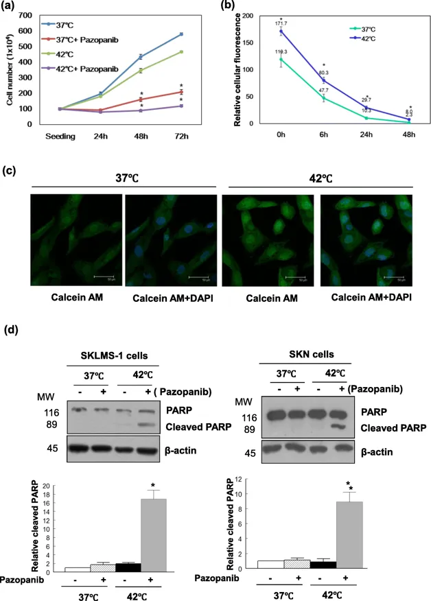
Pazopanib and Hyperthermia Synergistically Inhibit LMS Cell Proliferation by Inducing Apoptosis
Uterine leiomyosarcoma (LMS) is a rare, aggressive malignancy resistant to chemotherapy and radiotherapy. Pazopanib, a multitargeted tyrosine kinase inhibitor, is approved for advanced soft-tissue sarcomas but shows limited overall survival benefit. Regional hyperthermia (40-43°C) combined with chemotherapy reduces recurrence and mortality in high-risk soft-tissue sarcoma. Lin et al. investigated whether hyperthermia synergizes with pazopanib against LMS growth and explores underlying molecular mechanisms.
They initially identified the optimal hyperthermia protocol for uterine leiomyosarcoma (LMS) cells. Exposure to 42°C for 2 h did not induce cell death but maximized membrane permeability as shown by calcein AM assay. Dose-response curves for pazopanib under different conditions. SKLMS-1 cells treated with pazopanib and/or hyperthermia (42°C, 2 h) showed markedly reduced proliferation with combined treatment versus either alone (Fig. 1a). Hyperthermia alone increased calcein AM entry (Fig. 1b, c). Combined treatment synergistically induced apoptosis, evidenced by cleaved PARP levels exceeding the sum of individual treatments at 72 h in both SKLMS-1 and SKN cells (Fig. 1d). These data support apoptosis as the mechanism underlying pazopanib- and hyperthermia-induced LMS growth inhibition.
Ask a Question
Write your own review
- You May Also Need

Description: Uterine adenosquamous carcinoma. Said CEA and CA125 producing. Cell growth is slow.

Description: Established from tumor tissue from a 77-year-old woman with recurrence of endometrial carcinoma (adenomatous, partly papillary, grade G3) in 1990; described as forming heterotransplantable tumors in ...

Description: Glassy cell carcinoma. TA-4, CA125, neuron-specific enolase producing.
Description: The cells possess alpha keratin, well defined junctional complexes, tonofilaments and surface microvilli.

- Adipose Tissue-Derived Stem Cells
- Human Neurons
- Mouse Probe
- Whole Chromosome Painting Probes
- Hepatic Cells
- Renal Cells
- In Vitro ADME Kits
- Tissue Microarray
- Tissue Blocks
- Tissue Sections
- FFPE Cell Pellet
- Probe
- Centromere Probes
- Telomere Probes
- Satellite Enumeration Probes
- Subtelomere Specific Probes
- Bacterial Probes
- ISH/FISH Probes
- Exosome Isolation Kit
- Human Adult Stem Cells
- Mouse Stem Cells
- iPSCs
- Mouse Embryonic Stem Cells
- iPSC Differentiation Kits
- Mesenchymal Stem Cells
- Immortalized Human Cells
- Immortalized Murine Cells
- Cell Immortalization Kit
- Adipose Cells
- Cardiac Cells
- Dermal Cells
- Epidermal Cells
- Peripheral Blood Mononuclear Cells
- Umbilical Cord Cells
- Monkey Primary Cells
- Mouse Primary Cells
- Breast Tumor Cells
- Colorectal Tumor Cells
- Esophageal Tumor Cells
- Lung Tumor Cells
- Leukemia/Lymphoma/Myeloma Cells
- Ovarian Tumor Cells
- Pancreatic Tumor Cells
- Mouse Tumor Cells