
Human iPSC-derived Endothelial Cells
- Specification
- Background
- Scientific Data
- Q & A
- Customer Review
Human induced pluripotent stem cell-derived endothelial cells (iPSC-ECs) are generated by directed differentiation of hiPSCs through mesodermal and endothelial lineages using sequential growth factors and small molecules. They express canonical endothelial markers—CD31/PECAM-1, VE-cadherin, VEGFR2/KDR, CD34, von Willebrand factor, and eNOS—and exhibit endothelial functions including acetylated LDL uptake, UEA-1 lectin binding, migration, proliferation, tube formation, nitric oxide production, and barrier formation measurable by TEER/permeability.
Their key advantage is human, patient-specific origin with unlimited scalability. Unlike primary endothelial cells such as HUVECs, iPSC-ECs provide a renewable source, reduce donor variability through clonal selection, and enable repeated and high-throughput experiments. Because they retain the donor genotype, they are ideal for modeling genetic vascular diseases, diabetes, pulmonary hypertension, and retinal vasculopathies, and for personalized drug-response testing. Paired with CRISPR/Cas9, isogenic controls can be generated to dissect disease mechanisms. They also support co-culture with iPSC-derived cardiomyocytes, pericytes, or organoids and are explored for regenerative cell therapy and tissue engineering.
Crosstalk Between CGRP and Piezo1 in Human iPSC-Derived Endothelial Cells Relevant to Migraine
It is becoming increasingly evident that the vasculature is implicated in migraine pathophysiology. Calcitonin gene-related peptide (CGRP) acts as one of the key migraine mediators through various mechanisms that includes endothelium-mediated cerebral vessel vasodilation. Endothelial cells express mechanosensitive Piezo1 channels and have been suggested to play a role in migraine pathophysiology. However, the crosstalk between these two migraine-related signaling pathways remains unclear.
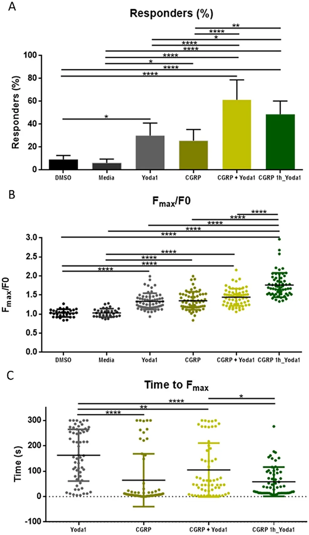
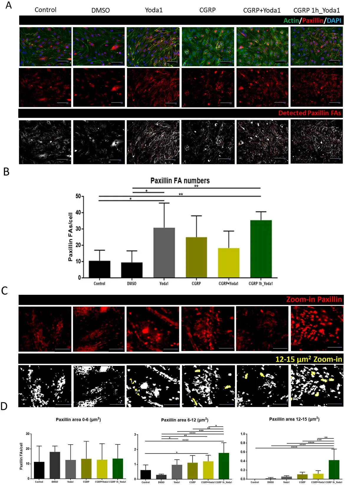
We measured intracellular calcium (Ca2+) in human induced pluripotent stem cell-derived endothelial cells (hiPSC-ECs), after exposure to Yoda1, a specific Piezo1 channel agonist, with and without CGRP. In addition, we investigated the effects of CGRP and Yoda1 on cellular remodeling by staining for focal adhesion (FA) protein paxillin using immunocytochemistry.
Our data suggest that a one-hour sensitization of hiPSC-ECs with CGRP followed by application of Yoda1 leads to a higher intracellular Ca2+ level compared to when Yoda1 and CGRP are acutely applied separately or combined, suggesting at least indirect crosstalk between the two signaling pathways in the vascular system. CGRP receptor antagonist BIBN4096 significantly reduced the intracellular Ca²+ level under this sensitization protocol, confirming effective CGRP pathway blockade. The results also show that a one-hour sensitization of CGRP and Piezo1 activation affects cellular remodeling as evidenced by an increased number and area size of paxillin FA points per cell in hiPSC-ECs.
Ask a Question
Write your own review
- Adipose Tissue-Derived Stem Cells
- Human Neurons
- Mouse Probe
- Whole Chromosome Painting Probes
- Hepatic Cells
- Renal Cells
- In Vitro ADME Kits
- Tissue Microarray
- Tissue Blocks
- Tissue Sections
- FFPE Cell Pellet
- Probe
- Centromere Probes
- Telomere Probes
- Satellite Enumeration Probes
- Subtelomere Specific Probes
- Bacterial Probes
- ISH/FISH Probes
- Exosome Isolation Kit
- Human Adult Stem Cells
- Mouse Stem Cells
- iPSCs
- Mouse Embryonic Stem Cells
- iPSC Differentiation Kits
- Mesenchymal Stem Cells
- Immortalized Human Cells
- Immortalized Murine Cells
- Cell Immortalization Kit
- Adipose Cells
- Cardiac Cells
- Dermal Cells
- Epidermal Cells
- Peripheral Blood Mononuclear Cells
- Umbilical Cord Cells
- Monkey Primary Cells
- Mouse Primary Cells
- Breast Tumor Cells
- Colorectal Tumor Cells
- Esophageal Tumor Cells
- Lung Tumor Cells
- Leukemia/Lymphoma/Myeloma Cells
- Ovarian Tumor Cells
- Pancreatic Tumor Cells
- Mouse Tumor Cells