
Human iPSC-derived Cortical Neurons
- Specification
- Scientific Data
- Q & A
- Customer Review
Circulating neuron-derived cfDNA for blood-based detection of Alzheimer’s and other neurodegenerative conditions
Background
This proof-of-concept study demonstrates that native nanopore sequencing can generate a human brain methylation atlas and classify neuron-derived cfDNA from blood plasma. Using six primary neural cell types, the authors developed cell-type-specific classifiers and applied them to 137 plasma samples from Alzheimer’s disease, Parkinson’s disease, ALS, mild cognitive impairment, and controls. Elevated cortical neuron-like cfDNA occurred in AD/MCI, dopaminergic neuron-like cfDNA in PD, and spinal motor neuron-like cfDNA in ALS. Multivariate integration improved diagnostic separation (AUC >0.85). Native sequencing avoids bisulfite/PCR bias, supporting liquid biopsy for neurodegenerative disease.
Key Findings
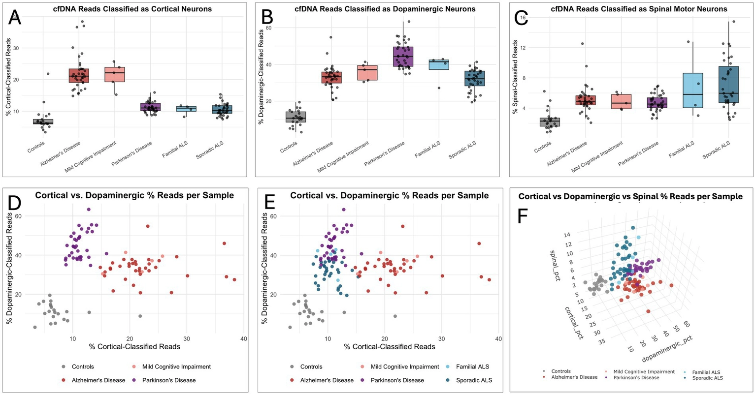
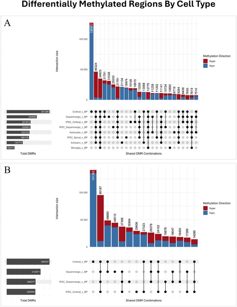
The study generated a methylation reference atlas from six primary human neural cell populations—cortical neurons, dopaminergic neurons, spinal motor neurons, astrocytes, Schwann cells, and microglia—using whole-genome native nanopore sequencing. This enabled cell-type-specific cfDNA classifiers with high analytical accuracy in in silico dilution series (R² up to 0.998). In 137 plasma samples, classifier signals aligned with selectively vulnerable neurons: cortical neuron-like cfDNA was elevated in AD and progressive MCI (22.27% vs 7.42% controls), dopaminergic neuron-like cfDNA in PD (45.01% vs 10.94%), and spinal motor neuron-like cfDNA in ALS (7.11% vs 2.48%). Multivariate integration of neuronal signatures improved disease discrimination, achieving AUC >0.85 in multi-disease comparisons. iPSC-derived neuron classifiers failed to replicate disease signals, highlighting the value of primary-cell references. The work supports native cfDNA nanopore methylation sequencing as a flexible platform for cell-of-origin analysis in neurodegeneration.
Contribution
Creative Bioarray supplied purified human neural cell populations, including primary and iPSC-derived cortical and dopaminergic neurons and glial cells, used to generate the whole-genome nanopore methylation reference atlas. These reference methylomes enabled development of cell-type-specific cfDNA classifiers for neurodegeneration analysis.
Circulating Cell-Free DNA Methylation Profiles Enable Disease-Specific Detection of Alzheimer’s, Parkinson’s, and ALS from Blood
Background
This study presents a blood-based cfDNA methylation platform for noninvasive detection of neurodegenerative diseases. Whole-genome nanopore methylation atlases of six primary human neural cell types were used to train classifiers assigning cfDNA to neuronal or glial origins. Classifiers were validated in silico and applied to 219 plasma samples from Alzheimer’s disease, Parkinson’s disease, amyotrophic lateral sclerosis, and controls. Disease-specific cfDNA elevations were detected, with predictive AUCs >0.99. In mild cognitive impairment, cortical cfDNA elevation identified individuals who later progressed to Alzheimer’s disease. A multivariate model improved accuracy, supporting cfDNA methylation profiling as a scalable framework.
Key Findings
The authors developed a cfDNA methylation platform capable of resolving cell-of-origin signals in blood. A whole-genome nanopore methylation atlas covering cortical, dopaminergic, and spinal motor neurons, astrocytes, Schwann cells, and microglia enabled classifiers that assign cfDNA to neural cell types. In 219 plasma samples, cortical cfDNA was elevated in Alzheimer’s disease, dopaminergic cfDNA in Parkinson’s disease, and spinal motor neuron cfDNA in amyotrophic lateral sclerosis, achieving AUCs >0.99. Mild cognitive impairment cases that later converted to Alzheimer’s disease showed elevated cortical cfDNA. Multivariate modeling integrating multiple neuronal signals improved sensitivity, specificity, and accuracy. The study demonstrates that cfDNA methylation profiling can detect and differentiate neurodegenerative diseases noninvasively, with potential for early detection and monitoring.
Contribution
Creative Bioarray offers purified human neurons and glial cells, including primary and iPSC-derived cortical neurons (CSC-C1514; CSC-00834L), primary and iPSC-dopaminergic neurons (CSC-C30012; CSC-00842L), as well as human astrocytes (CSC-7836W), human Schwann cells (CSC-7836W) and human microglia (CSC-C1528), which are used to construct a whole-genome nanopore methylation reference map and train a classifier for cell source classification from cfDNA.
Ask a Question
Write your own review
- Adipose Tissue-Derived Stem Cells
- Human Neurons
- Mouse Probe
- Whole Chromosome Painting Probes
- Hepatic Cells
- Renal Cells
- In Vitro ADME Kits
- Tissue Microarray
- Tissue Blocks
- Tissue Sections
- FFPE Cell Pellet
- Probe
- Centromere Probes
- Telomere Probes
- Satellite Enumeration Probes
- Subtelomere Specific Probes
- Bacterial Probes
- ISH/FISH Probes
- Exosome Isolation Kit
- Human Adult Stem Cells
- Mouse Stem Cells
- iPSCs
- Mouse Embryonic Stem Cells
- iPSC Differentiation Kits
- Mesenchymal Stem Cells
- Immortalized Human Cells
- Immortalized Murine Cells
- Cell Immortalization Kit
- Adipose Cells
- Cardiac Cells
- Dermal Cells
- Epidermal Cells
- Peripheral Blood Mononuclear Cells
- Umbilical Cord Cells
- Monkey Primary Cells
- Mouse Primary Cells
- Breast Tumor Cells
- Colorectal Tumor Cells
- Esophageal Tumor Cells
- Lung Tumor Cells
- Leukemia/Lymphoma/Myeloma Cells
- Ovarian Tumor Cells
- Pancreatic Tumor Cells
- Mouse Tumor Cells