
HMV-II
Cat.No.: CSC-C6251J
Species: Homo sapiens (Human)
Source: Vagina
Morphology: epithelial-like
Culture Properties: Adherent cells
- Specification
- Background
- Scientific Data
- Q & A
- Customer Review
Store in liquid nitrogen.
HMV-II (IIIB) cell line is a human malignant melanoma cell line originally derived from a metastatic lesion. It is commonly used as an in vitro model of melanoma tumor progression. These cells are able to form adherent monolayer cultures and demonstrate fibroblast-like morphology. They are rapidly proliferating and show stable growth. HMV-II cells have been shown to express melanocytic markers such as tyrosinase and MITF. HMV-II melanoma cells show increased melanogenesis.
HMV-II cell lines express dysregulated pathways associated with cell cycle regulation, cell survival and cell migration which are commonly seen in malignant melanomas. This line has been used in research to study melanoma invasion and metastasis as well as resistance to anti-cancer drugs. Furthermore, HMV-II has been used to screen anti-cancer drugs such as targeted therapeutics and combination treatments.
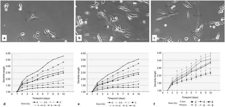
Vaginal Mucosal Melanoma Cell Activation in Response to Photon or Carbon Ion Irradiation
Primary gynecological melanomas are rare and more lethal than cutaneous forms; whether mucosal melanocytes modulate radiosensitivity is unknown. Charalampopoulou et al. examined how photon vs carbon-ion irradiation affects morphology, melanogenesis and motility of human vaginal-mucosal HMV-II melanoma cells.
Under standard culture conditions, HMV-II melanoma cells maintained epithelial morphology with triangular or spindle-shaped bodies and short processes. Following XRT or CIRT exposure, cells exhibited dose- and radiation-type-dependent morphological activation characterized by dendrite formation and elongated morphology (Fig. 1a-c). After photon irradiation, average dendrite length increased progressively with dose, peaking at 6 Gy and declining at 8-10 Gy by 24 h; this morphology persisted for 10 days (Fig. 1d). Carbon ion irradiation produced less pronounced changes at 24 h, but dendrite elongation emerged by 48 h, similarly peaking at 6 Gy and maintaining this pattern through day 10 (Fig. 1e). Notably, XRT induced significantly greater dendrite elongation than CIRT at all time points (Fig. 1f).
Ask a Question
Write your own review
- You May Also Need

Description: Uterine adenosquamous carcinoma. Said CEA and CA125 producing. Cell growth is slow.

Description: Established from tumor tissue from a 77-year-old woman with recurrence of endometrial carcinoma (adenomatous, partly papillary, grade G3) in 1990; described as forming heterotransplantable tumors in ...

Description: Glassy cell carcinoma. TA-4, CA125, neuron-specific enolase producing.
Description: The cells possess alpha keratin, well defined junctional complexes, tonofilaments and surface microvilli.

- Adipose Tissue-Derived Stem Cells
- Human Neurons
- Mouse Probe
- Whole Chromosome Painting Probes
- Hepatic Cells
- Renal Cells
- In Vitro ADME Kits
- Tissue Microarray
- Tissue Blocks
- Tissue Sections
- FFPE Cell Pellet
- Probe
- Centromere Probes
- Telomere Probes
- Satellite Enumeration Probes
- Subtelomere Specific Probes
- Bacterial Probes
- ISH/FISH Probes
- Exosome Isolation Kit
- Human Adult Stem Cells
- Mouse Stem Cells
- iPSCs
- Mouse Embryonic Stem Cells
- iPSC Differentiation Kits
- Mesenchymal Stem Cells
- Immortalized Human Cells
- Immortalized Murine Cells
- Cell Immortalization Kit
- Adipose Cells
- Cardiac Cells
- Dermal Cells
- Epidermal Cells
- Peripheral Blood Mononuclear Cells
- Umbilical Cord Cells
- Monkey Primary Cells
- Mouse Primary Cells
- Breast Tumor Cells
- Colorectal Tumor Cells
- Esophageal Tumor Cells
- Lung Tumor Cells
- Leukemia/Lymphoma/Myeloma Cells
- Ovarian Tumor Cells
- Pancreatic Tumor Cells
- Mouse Tumor Cells