
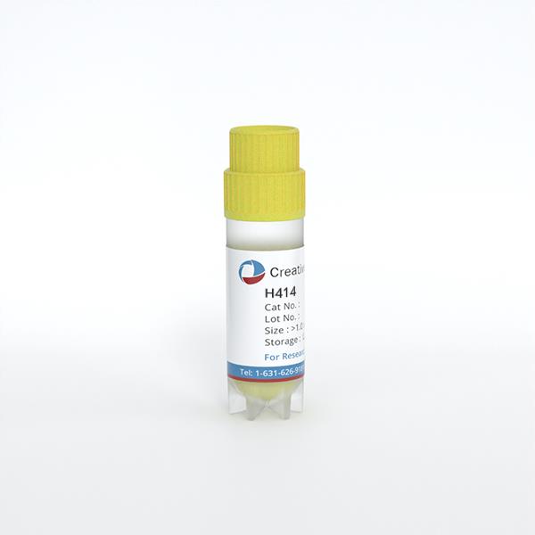
H414
Cat.No.: CSC-C6257J
Species: Homo sapiens (Human)
Source: Intestine; Colon
Morphology: epithelial-like
Culture Properties: Adherent cells
- Specification
- Background
- Scientific Data
- Q & A
- Customer Review
Store in liquid nitrogen.
The H414 (H-414) cell line is a human colorectal cancer-derived epithelial cell type frequently employed in research of DNA mismatch repair (MMR) and genome stability. It is a derivation or closely related subline of the HCT116 colorectal cancer cell system and is particularly useful for comparative functional studies of MMR-proficient and MMR-deficient backgrounds.
In contrast to the parental HCT116 cells with no mismatch repair (MMR) activity due to defective MLH1 expression, H414 cells have functional MMR activity and serve as an important parallel model for DNA repair studies. This enhanced repair capacity allows researchers to explore the effects of MMR proficiency on mutagenesis, microsatellite stability, and sensitivity to DNA damaging agents.
H414 cells have epithelial-like shape and develop as adherent monolayers under conventional culture conditions. They are widely employed in cancer biology, pharmacology and molecular genetics research, in especially in the evaluation of chemotherapeutic drugs such as 5-fluorouracil (5-FU) and other DNA-damaging chemicals. Due to their stable genetic background, they are also appropriate for studies involving gene editing, DNA damage response pathways and cell cycle regulation.
Ganciclovir Triphosphate Incorporation Drives Mutagenesis in Hematopoietic and Colorectal Cells
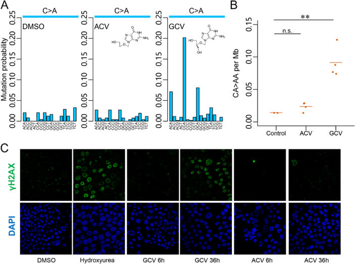
Ganciclovir (GCV) is a mainstay for cytomegalovirus management in transplant patients, yet its potential mutagenicity remains poorly defined. Clinical review of two hematopoietic stem cell transplant (HSCT) patients who developed colorectal neoplasms harboring the GCV-associated mutational signature (GCVsig) revealed prior exposure to both GCV and acyclovir (ACV). To determine the specificity of GCVsig, Fang et al. treated the H414 colorectal cancer cell line with DMSO, GCV, or ACV. While cells tolerated treatment at 100 µM, only GCV exposure induced the characteristic CA > AA mutational signature, which was absent in ACV-treated samples (Fig. 1A). This specificity was validated in HoxA9-Meis1-transformed murine myeloid cells, which were less tolerant to antivirals. Despite a 20-fold lower dose (5 µM), only GCV-treated cells acquired significant CA > AA mutations compared to ACV or controls (p< 0.01, Student’s t-test) (Fig. 1B).
Mechanistically, GCV-triphosphate (GCV-TP) incorporation into DNA was confirmed by increased staining of the DNA damage marker γH2AX in H414 cells after 36 hours of GCV treatment—coinciding with DNA replication—whereas ACV-treated cells showed minimal γH2AX signal (Fig. 1C). Flow cytometry revealed no significant cell cycle arrest in either treatment group at 36 hours, indicating that GCV-induced mutagenesis occurs without overt cytotoxicity in tolerant cells.
Ask a Question
Write your own review
- You May Also Need
Description: Established from an adenocarcinoma located in the ascending colon of a 68 year-old male patient. The adenocarcinoma was well differentiated and determined to be Dukes' stage B. Imperial College ...
Description: This is one cell line out of a series of colon carcinoma cell lines established by PD Dr. Michael Linnebacher.
Description: Rectal carcinoma from a Japanese patient. Cell growth is slow.
Description: The C80 cell line was established from a 69-year old male patient with a moderately well differentiated adenocarcinoma of the rectum classified as Dukes' stage D.
Description: RKO is a poorly differentiated colon carcinoma cell line developed by Michael Brattain.
- Adipose Tissue-Derived Stem Cells
- Human Neurons
- Mouse Probe
- Whole Chromosome Painting Probes
- Hepatic Cells
- Renal Cells
- In Vitro ADME Kits
- Tissue Microarray
- Tissue Blocks
- Tissue Sections
- FFPE Cell Pellet
- Probe
- Centromere Probes
- Telomere Probes
- Satellite Enumeration Probes
- Subtelomere Specific Probes
- Bacterial Probes
- ISH/FISH Probes
- Exosome Isolation Kit
- Human Adult Stem Cells
- Mouse Stem Cells
- iPSCs
- Mouse Embryonic Stem Cells
- iPSC Differentiation Kits
- Mesenchymal Stem Cells
- Immortalized Human Cells
- Immortalized Murine Cells
- Cell Immortalization Kit
- Adipose Cells
- Cardiac Cells
- Dermal Cells
- Epidermal Cells
- Peripheral Blood Mononuclear Cells
- Umbilical Cord Cells
- Monkey Primary Cells
- Mouse Primary Cells
- Breast Tumor Cells
- Colorectal Tumor Cells
- Esophageal Tumor Cells
- Lung Tumor Cells
- Leukemia/Lymphoma/Myeloma Cells
- Ovarian Tumor Cells
- Pancreatic Tumor Cells
- Mouse Tumor Cells