
WCP 12 FISH Probe
- Specification
- Scientific Data
- Q & A
- Customer Review
- Documents
 For Research Use Only. Not for use in diagnostic procedures.
Characterization of Human Herpesvirus 8 genomic integration and amplification events in a primary effusion lymphoma cell line
Background
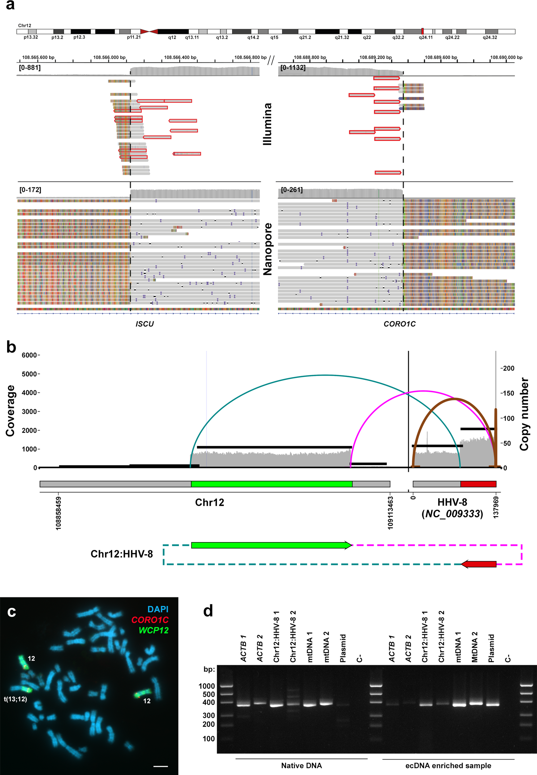
This study investigated Human Herpesvirus 8 (HHV-8) integration into the human genome in the primary effusion lymphoma cell line BC-3 using short- and long-read sequencing. Two highly supported HHV-8 integrants were identified on chromosome 12, encompassing a ~1.2 Mb region amplified ~16-fold. The event may represent the first known HHV-8 integration into hybrid human-viral extrachromosomal chimeric circular DNA (eccDNA). The amplified region included SELPLG and CORO1C. Nanopore methylation analysis indicated the SELPLG promoter CpGs were mostly unmethylated, suggesting transcriptional activity. These findings implicate viral insertion and eccDNA amplification in HHV-8-associated oncogenesis.
Key Findings
The study identified two robust HHV-8-human junctions on chromosome 12 in BC-3 cells, supported by independent NGS and Nanopore runs. These junctions flanked an approximately 1.2 Mb region amplified ~16-fold, containing partial ISCU, complete SELPLG and TMEM119, and truncated CORO1C. Nanopore assemblies recovered ~30 Kb of HHV-8 and up to 16 LTR repeats, suggesting a single integration event. AmpliconArchitect indicated a bifocal amplicon linking chr12 to HHV-8, consistent with an extrachromosomal circular DNA (ecDNA/eccDNA) configuration. PCR after exonuclease V enrichment confirmed the circular nature of these chimeric HHV-8-Chr12 DNAs. Methylation analysis showed the SELPLG promoter CpGs were predominantly unmethylated (96.774%), implying the amplified SELPLG copies are transcriptionally active. FISH and PRINS were inconclusive for extrachromosomal localization. The authors propose that viral insertion followed by eccDNA amplification may be a mechanism in HHV-8-associated cancers, and stress validation with multiple sequencing approaches.
Contribution
Creative Bioarray provided the commercial whole chromosome 12 paint probe (FWCP-12) used in dual FISH experiments on BC-3 metaphase spreads. This probe enabled visualization of chromosome 12, helping assess Chr12q aneuploidy and cytogenetically characterize the HHV-8–Chr12 rearrangement in the study.
Ask a Question
Write your own review
 Datasheet
- Adipose Tissue-Derived Stem Cells
- Human Neurons
- Mouse Probe
- Whole Chromosome Painting Probes
- Hepatic Cells
- Renal Cells
- In Vitro ADME Kits
- Tissue Microarray
- Tissue Blocks
- Tissue Sections
- FFPE Cell Pellet
- Probe
- Centromere Probes
- Telomere Probes
- Satellite Enumeration Probes
- Subtelomere Specific Probes
- Bacterial Probes
- ISH/FISH Probes
- Exosome Isolation Kit
- Human Adult Stem Cells
- Mouse Stem Cells
- iPSCs
- Mouse Embryonic Stem Cells
- iPSC Differentiation Kits
- Mesenchymal Stem Cells
- Immortalized Human Cells
- Immortalized Murine Cells
- Cell Immortalization Kit
- Adipose Cells
- Cardiac Cells
- Dermal Cells
- Epidermal Cells
- Peripheral Blood Mononuclear Cells
- Umbilical Cord Cells
- Monkey Primary Cells
- Mouse Primary Cells
- Breast Tumor Cells
- Colorectal Tumor Cells
- Esophageal Tumor Cells
- Lung Tumor Cells
- Leukemia/Lymphoma/Myeloma Cells
- Ovarian Tumor Cells
- Pancreatic Tumor Cells
- Mouse Tumor Cells