
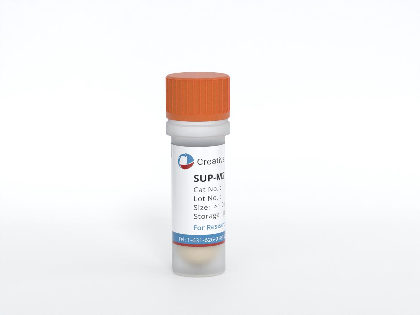
SUP-M2
Cat.No.: CSC-C0518
Species: Homo sapiens (Human)
Source: Cerebrospinal Fluid
Morphology: round cells growing singly or in clumps in suspension
Culture Properties: suspension
- Specification
- Background
- Scientific Data
- Q & A
- Customer Review
- Documents
Immunology: CD2 +, CD3 -, CD4 +, CD13 -, CD14 -, CD19 -, CD21 -, CD25 +, CD30 +, CD34 -, CD71 +, HLA-DR +
Viruses: PCR: EBV -, HBV -, HCV -, HIV -, HTLV-I/II -, SMRV -
SUP-M2 cells are a human cell line derived from the cerebrospinal fluid of a 5-year-old girl with refractory malignant histiocytosis. These cells are characterized by specific genetic and phenotypic features, making them an important model for studying a particular type of cancer known as anaplastic large cell lymphoma (ALCL).
The SUP-M2 cell line is notable for carrying the characteristic translocation t (2; 5) (p23; q35). This translocation leads to the fusion of two genes, NPM1 and ALK, resulting in the formation of the NPM1-ALK fusion gene. The NPM1-ALK fusion gene is a hallmark genetic alteration in a subset of ALCL cases and plays a crucial role in the development and progression of this disease.
In addition to the genetic abnormality, SUP-M2 cells also express the CD30 antigen. CD30 is a cell surface marker that is commonly observed on ALCL cells. The presence of CD30 further defines SUP-M2 cells as anaplastic large-cell lymphoma and contributes to their phenotypic characteristics.
The SUP-M2 cell line has been widely used in research to investigate the biological and molecular aspects of ALCL, including the role of the NPM1-ALK fusion gene, cellular signaling pathways, therapeutic targets, and drug testing. By studying SUP-M2 cells, researchers aim to gain insights into the pathogenesis of ALCL and develop more effective treatment strategies for this type of lymphoma.
Super enhancer acquisition drives expression of oncogenic PPP1R15B that regulates protein homeostasis in multiple myeloma
Background
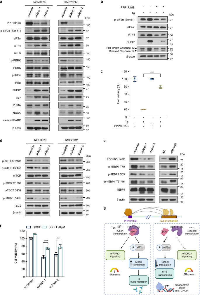
Multiple myeloma (MM) is a hematological malignancy with poorly understood chromatin rewiring. This study integrates H3K27ac ChIP-seq and HiChIP to map myeloma-associated super-enhancers (SEs). It identifies PPP1R15B as an SE-driven oncogene encoding a regulatory subunit of the eIF2α phosphatase complex. PPP1R15B silencing activates the pro-apoptotic eIF2α-ATF4-CHOP pathway and inhibits protein synthesis and immunoglobulin production. Pharmacological inhibition of PPP1R15B with Raphin1 potentiates bortezomib's anti-myeloma effect, highlighting a promising therapeutic strategy targeting protein homeostasis.
Key Findings
Integrative analysis of H3K27ac ChIP-seq and HiChIP identified 223 gained super-enhancers (SEs) in multiple myeloma (MM), leading to a prioritized list of nine SE-associated genes. PPP1R15B was found to be overexpressed in MM and associated with poor survival. Its SE is activated by transcription factors CEBPB and YY1. PPP1R15B depletion inhibited cell proliferation, induced apoptosis, and suppressed global protein synthesis and immunoglobulin secretion. Mechanistically, PPP1R15B dephosphorylates eIF2α, suppressing pro-apoptotic UPR and activating mTORC1 signaling. In vivo, PPP1R15B knockdown prolonged survival in a xenograft model. Pharmacological inhibition with Raphin1 induced apoptosis in MM cells and synergized with bortezomib. These findings demonstrate that PPP1R15B is a critical regulator of protein homeostasis and a promising therapeutic target in MM.
Contribution
Creative Bioarray provided the anaplastic large cell lymphoma cell line SUP-M2 (Cat# CSC-C0518), which was used to assess the therapeutic potential of PPP1R15B inhibition in other cancer types. This contributed to demonstrating the broader applicability of targeting PPP1R15B beyond multiple myeloma.
B7-H3 Redirected CAR-T Cells Control ALCLs In Vitro
B7 homolog 3 (B7-H3), also named CD276, is a member of the B7 family of immune regulatory proteins. B7-H3 CAR-T cells showed strong cytotoxicity and cytokine secretion against target ALCL cells (SUP-M2, SU-DHL-1, and Karpas 299) in vitro. Cytotoxicity was measured using two different approaches LDH release cytotoxic assay and luciferase-based assay in various E: T ratios. Increased cytotoxicity along with an increased E: T ratio was observed in B7-H3 CAR-T cells to target ALCL cell lines in both approaches. Furthermore, >60% specific lysis was reached when the E: T ratio was 5:1 for all targets in the LDH release cytotoxic assay (Fig. 1A).
To confirm this effect, stable expressing luciferase cell lines were generated from parental SUP-M2, Karpas299, and SU-DHL-1 cell lines as well as the performed luciferase-based assay. This was consistent with data from the LDH release cytotoxic assay, and B7-H3 CAR-T cells showed their potency in controlling SUP-M2, Karpas299, and SU-DHL-1 (Fig. 1B). The secreting of IL-2 and IFNγ was observed at a high level when B7-H3 T cells were co-cultured with ALCLs. In contrast, UTD and CD19 CAR-T control cells showed minimal secretion of both cytokines (Fig. 1C and D).
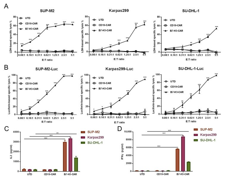 Fig. 2. B7-H3 T cell effects on B7-H3-expressing ALCL cells (SUP-M2, SU-DHL-1, and Karpas 299). (Slupianek A and Skorski T, 2004)
Ask a Question
Write your own review
 SUP-M2
- You May Also Need
Description: Established in 2007 from the bone marrow mononuclear cells of an 82-year-old Japanese man with diffuse large B-cell lymphoma in the leukemic phase
Description: The MEG-01 cell line was derived in 1983 at the Nagoya University School of Medicine, Nagoya, Japan from bone marrow cells taken from a patient in megakaryoblastic crisis of CML
Description: Established from the bone marrow of a 28-year-old man who developed the terminal leukemic phase of lymphosarcoma in 1976
Description: This cell line was derived from the bone marrow aspirate of a 59 year old male with erythroleukemia that became acute myelogenous leukaemia.The cells form colonies in soft-agar in the presence of ...
Description: Established from the pleural effusion of a 24-year-old woman with recurrent anaplastic large cell lymphoma (ALCL); cells were described to clonally derive from T-lineage lymphoid cells and to be ...
Description: Established from a 37-year-old man at second (refractory/terminal) relapse of Hodgkin lymphoma (nodular sclerosing -> lymphocyte depleted/stage IIISA -> stage IV) after both combined chemo- and ...
- Adipose Tissue-Derived Stem Cells
- Human Neurons
- Mouse Probe
- Whole Chromosome Painting Probes
- Hepatic Cells
- Renal Cells
- In Vitro ADME Kits
- Tissue Microarray
- Tissue Blocks
- Tissue Sections
- FFPE Cell Pellet
- Probe
- Centromere Probes
- Telomere Probes
- Satellite Enumeration Probes
- Subtelomere Specific Probes
- Bacterial Probes
- ISH/FISH Probes
- Exosome Isolation Kit
- Human Adult Stem Cells
- Mouse Stem Cells
- iPSCs
- Mouse Embryonic Stem Cells
- iPSC Differentiation Kits
- Mesenchymal Stem Cells
- Immortalized Human Cells
- Immortalized Murine Cells
- Cell Immortalization Kit
- Adipose Cells
- Cardiac Cells
- Dermal Cells
- Epidermal Cells
- Peripheral Blood Mononuclear Cells
- Umbilical Cord Cells
- Monkey Primary Cells
- Mouse Primary Cells
- Breast Tumor Cells
- Colorectal Tumor Cells
- Esophageal Tumor Cells
- Lung Tumor Cells
- Leukemia/Lymphoma/Myeloma Cells
- Ovarian Tumor Cells
- Pancreatic Tumor Cells
- Mouse Tumor Cells