
Rat Bone Marrow Mesenchymal Stem Cells
- Specification
- Background
- Scientific Data
- Q & A
- Customer Review
Rat Bone Marrow Mesenchymal StemCells are negative for bacteria, yeast, fungi, and mycoplasma. Cells can be expanded on a multiwell culture plate ready for experiments under the cell culture conditions specified by Creative Bioarray. Repeated freezing and thawing of cells is not recommended.
Mouse Bone Marrow Mesenchymal Stem Cells are tested for expression of markers using antibodies, CD44, Sca-1 and CD29 by flow cytometry.
Standard biochemical procedures performed include the assays of cell to cell interaction, PCR, Western blotting, immunoprecipitation, immunofluorescent staining, immunofluorescent flow cytometry or generating cell derivatives for desired research applications.
Rat Bone Marrow Mesenchymal Stem Cells (BM-MSCs), also termed bone marrow-derived mesenchymal stromal cells, are nonhematopoietic stromal populations isolated from rat bone marrow. Under appropriate culture conditions, they exhibit plastic adherence, fibroblast-like morphology, and the capacity for osteogenic, adipogenic, and chondrogenic differentiation. These characteristics make them a versatile model for investigating mesenchymal lineage commitment, stromal biology, and mechanisms relevant to tissue regeneration.
A principal advantage is their capacity for culture expansion, enabling sufficient cell numbers for complementary molecular and functional experiments. Their differentiation potential supports controlled studies of extracellular matrix production, mineralization, lipid accumulation, and cartilage-associated matrix formation.
Beyond differentiation, these cells provide a platform for investigating paracrine signaling and immunomodulatory interactions. Rat BM-MSCs secrete bioactive factors whose abundance and activity can change with environmental stimulation. Conditioned-medium studies have demonstrated effects on neurite outgrowth and vascular smooth muscle cell behavior, supporting investigation of mechanisms that do not require direct differentiation into replacement tissue. This functional flexibility enables studies of cellular communication, preconditioning strategies, and factors contributing to repair responses.
Their compatibility with established rat experimental models further facilitates comparisons between in vitro findings and tissue-level outcomes. Applications include biomaterial evaluation, bone regeneration research, and analysis of immune–stromal interactions.
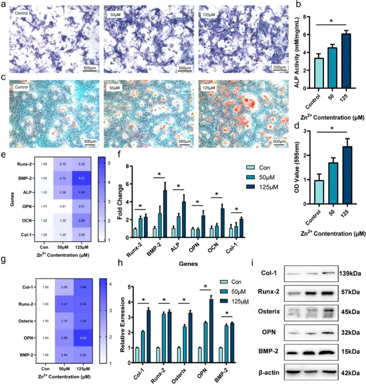
Twofold Effects of Zn2+ on Bone Marrow Mesenchymal Stem Cells—Osteogenesis or Apoptosis
Zinc (Zn) is a bioabsorbable metal that shows great potential as an implant material for orthopedic applications. Suitable concentrations of zinc ions promote osteogenesis, while excess zinc ions cause apoptosis. As a result, the conflicting impacts of Zn2+ concentration on osteogenesis could prove to be significant problems for the creation of novel materials.
This study thoroughly examined the cell viability, proliferation, and osteogenic differentiation of rat bone marrow mesenchymal stem cells (rBMSCs) cultured in various concentrations of Zn2+ in vitro. The effective promotion of cell survival, proliferation, migration, and osteogenic differentiation of bone marrow mesenchymal stem cell (BMSCs) may be achieved at a low concentration of Zn2+ (125 μM). The excessively high concentration of zinc ions (>250 μM) not only reduces BMSCs’ viability and proliferation but also causes them to suffer apoptosis due to the disturbed zinc homeostasis and excessive Zn2+. Moreover, transcriptome sequencing was used to examine the underlying mechanisms of zinc-induced osteogenic differentiation with particular attention paid to the PI3K-AKT and TGF-β pathways. The present investigation elucidated the dual impacts of Zn2+ microenvironments on the osteogenic characteristics of rBMSCs and might offer significant insights for refining the blueprint for zinc-based biomaterials.
Ask a Question
Write your own review
- You May Also Need
- Adipose Tissue-Derived Stem Cells
- Human Neurons
- Mouse Probe
- Whole Chromosome Painting Probes
- Hepatic Cells
- Renal Cells
- In Vitro ADME Kits
- Tissue Microarray
- Tissue Blocks
- Tissue Sections
- FFPE Cell Pellet
- Probe
- Centromere Probes
- Telomere Probes
- Satellite Enumeration Probes
- Subtelomere Specific Probes
- Bacterial Probes
- ISH/FISH Probes
- Exosome Isolation Kit
- Human Adult Stem Cells
- Mouse Stem Cells
- iPSCs
- Mouse Embryonic Stem Cells
- iPSC Differentiation Kits
- Mesenchymal Stem Cells
- Immortalized Human Cells
- Immortalized Murine Cells
- Cell Immortalization Kit
- Adipose Cells
- Cardiac Cells
- Dermal Cells
- Epidermal Cells
- Peripheral Blood Mononuclear Cells
- Umbilical Cord Cells
- Monkey Primary Cells
- Mouse Primary Cells
- Breast Tumor Cells
- Colorectal Tumor Cells
- Esophageal Tumor Cells
- Lung Tumor Cells
- Leukemia/Lymphoma/Myeloma Cells
- Ovarian Tumor Cells
- Pancreatic Tumor Cells
- Mouse Tumor Cells